


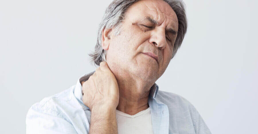
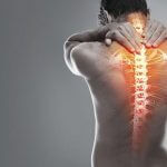
Neck Pain
Arched spine is composed of four parts as follows:
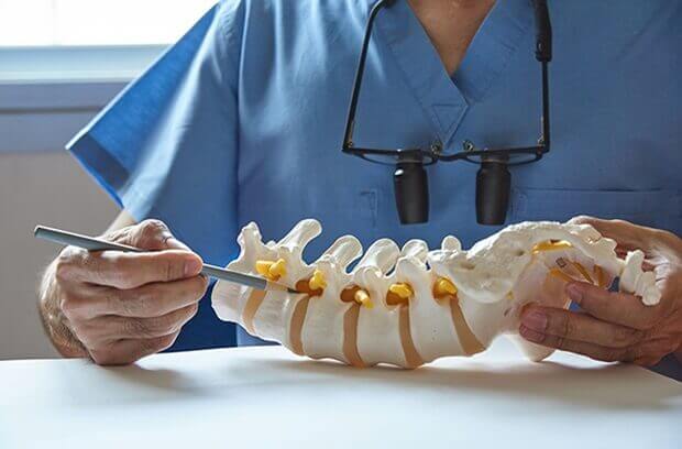
The upper arch is located at the head, neck or cervical Cervical arc and its convexity towards the front of the arch is composed of 7 pieces. The spine from top to bottom and from one to seven are named as C1-C7
The seven vertebrae of the spine are the thirty-three.
The first vertebra of the neck vertebrae, the atlas is called a vertebral body is not a bony ring. Axxis the second nut that connects to the spine, causing the motor to rotate the left and right. Seventh cervical vertebra with spinal processes and the long term is more than the rest of the cervical spine. Is palpable in the posterior neck and cervical spine or back is also distinctive.
● Neck Anatomy
A flexible region of the spine, the neck (cervical), respectively. Intervertebral disc consists of seven vertebrae of the cervical spine. The weight of the nut, the various movements that are. Nerve roots of the spine in the neck and upper limbs crossed, then the arms, forearms and fingers along the rip. There are many muscles in the neck. Some of the occipital Thoracic vertebrae have been drawn up.
Spine, the spine is made up of tandem. Every piece of bone, called the vertebral body consists of a cylindrical low in the back of the ring
Exposed bone (the first and second cervical vertebrae are just a ring). Placing the ring on the bony vertebral bone is largely a pipe that
Seat and protects the spinal cord.
Two sides of the spinal cord in the spinal nerve roots exit the spinal cord through a hole called the foramen Foramen out.
Inter-vertebral disc tissue is low cylindrical shape made of cartilage-like sex. Disk is located between the vertebrae, elasticity and
Facilitate and regulate their movements.
Disc is composed of two parts. A peripheral portion and a central portion. The outer part of which forms a ring called the annulus Fybrvzvs Anulus fibrosus
Thick and sturdy but flexible like rubber. Located within the central part of the environment and is soft and gelatinous. The central part of the nucleous Pvlpvzvs
Nucleus pulposus say the main character drives the shock and impact absorption for the same parts.
Age, trauma, or diseases such as arthritis, poor stature can lead to degeneration of the bones and joints of the cervical spine, causing disc herniation or event
The bone spur. In addition, sudden severe injuries to the neck may be a herniated disc, neck strain, damage to blood vessels, bones and bone damage
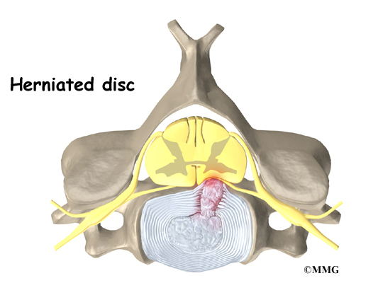
Or ligament, and in extreme cases, lead to permanent paralysis. After getting into too much pressure on the spine, peripheral annular portion of the disc (annulus Fybrvzvs) and the ruptured
Central (nucleous Pvlpvzvs) the gap is torn out of the way. This part of the hernia protruding or herniated say. The main problem with disc herniation occurs
That the protruding part of the nerve roots and spinal cord that are coming out of the pressure. Contact the chemical constituents of the gelatinous
Disc with nerve root irritation and inflammation caused them. The pressure and nerve inflammation causing clinical symptoms in these patients.
The incidence of herniation or disc herniation in the neck below the waist. Due to its special structure and the strength of the ligament in the cervical spine, the vertebrae and discs
Communication, protection and adds interest to the muscles, bones, cartilage, blood vessels and nerves in the spine and cervical spine with each other, a disk with neck pain
Arms and neck begins, and thus the effect of pressure vessels, nerves and tissues surrounding the spinal disc is inserted, there is also the possibility of numbness and downstream
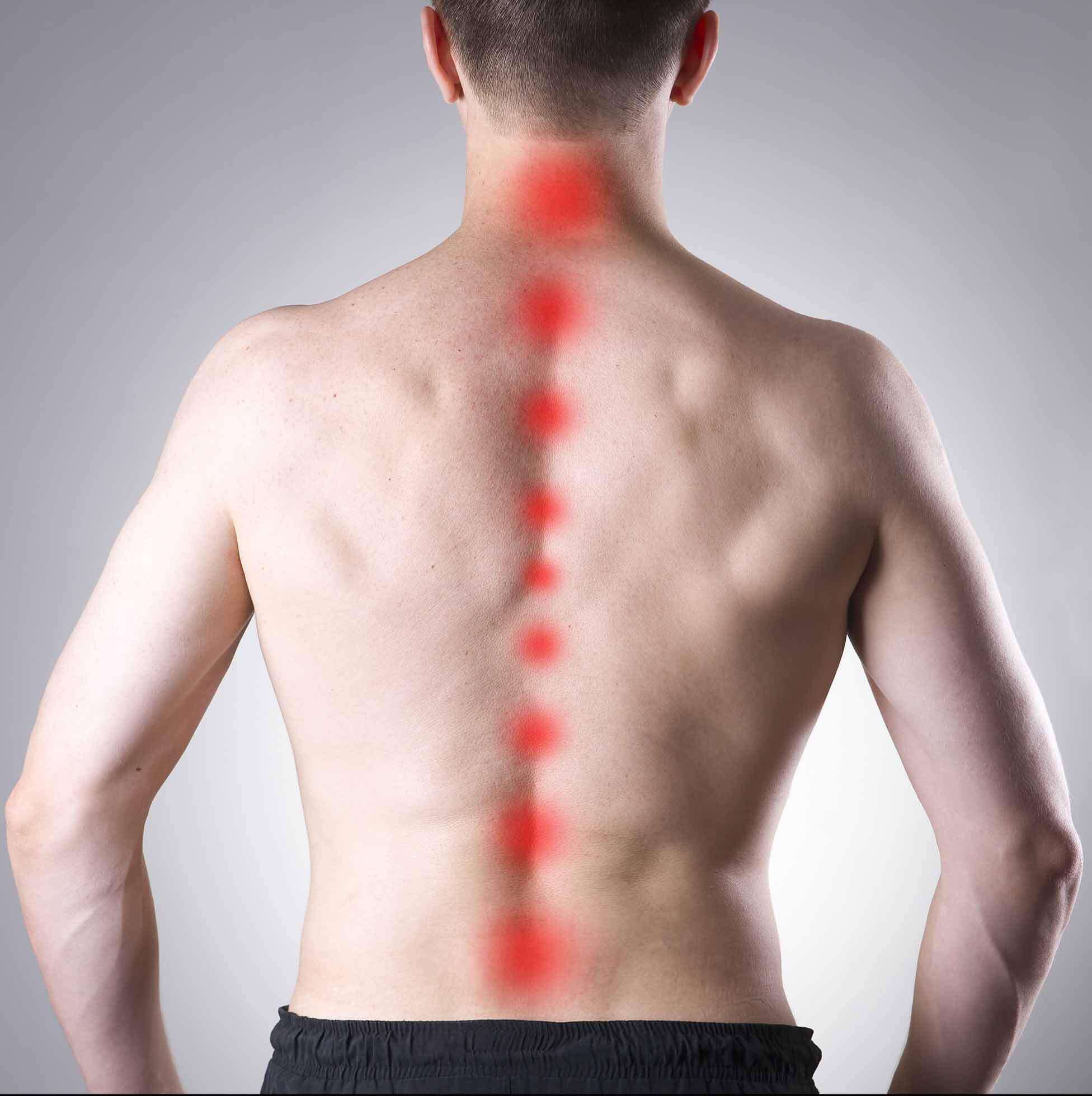
Even the fingertips, we Hychh weakening of the neck, the most common cervical vertebrae in the spine hurt five, six and seven in the lower part of the neck
Located, in case of damage to the spinal nerve roots, four and five of the fifth vertebra pain in the shoulder of the person reflected in the fifth and sixth vertebrae damage
Cervical spinal nerve root pain into our sixth Hychh guided by the hand and wrist, and weaken them, are the most common type of cervical disc
Is, if they are damaged, sixth and seventh vertebrae, spinal nerve root pain seventh Hychh back to our side of the hand and guided along the front of the
Track, and weaken them, causing numbness in the fingertips, which also shows
Or herniated cervical disc herniation occurs in young females. The prevalence of smokers among men and especially more frequent and more disk cervical vertebrae 5 and 6 and in
The spinal cord is a column of nerve tissue protected by a bony tube in the spinal column. Conditions that narrow the space in this tube put the spinal cord at risk of getting squeezed. This narrowing in the spinal column of the neck is called cervical spinal stenosis, or cervical stenosis. Pressure against the spinal cord as a result of spinal stenosis causes myelopathy, a condition that demands medical attention. Myelopathy can cause problems with the bowels and bladder, change the way you walk, and affect your ability to use your fingers and hands.
Anatomy
What parts make up the spine and neck?
The spine is made of a column of bones. Each bone, or vertebra, is formed by a round block of bone, called a vertebral body. A bony ring attaches to the back of the vertebral body, forming a canal.
This bony ring is formed by two sets of bones. One set, the pedicle bones, attaches to the back of each vertebral body. On the other end, each pedicle bone connects with a lamina bone. The lamina bones form a protective roof over the back of the spinal cord. When the vertebra bones are stacked on top of each other, the bony rings forms a long bony tube that surrounds and protects the spinal cord as it passes through the spine.
An intervertebral disc fits between each vertebral body and provides a space between the spine bones. The disc works like a shock absorber. It protects the spine against the daily pull of gravity. It also protects the spine during activities that put strong force on the spine, such as jumping, running, and lifting.
An intervertebral disc is made up of two parts. The center, called the nucleus, is spongy. It provides most of the ability to absorb shock. The nucleus is held in place by the annulus, a series of strong ligament rings surrounding it. Ligaments are strong connective tissues that attach bones to other bones.
Causes
Why do I have this problem?
The bony spinal canal normally has more than enough room for the spinal cord. Typically, the canal is 17 to 18 millimeters around, slightly less than the size of a penny. Spinal stenosis occurs when the canal narrows to 13 millimeters or less. When the size drops to 10 millimeters, severe symptoms of myelopathy occur. Myelopathy is a term for any condition that affects the spinal cord. The symptoms of myelopathy result from pressure against the spinal cord and reduced blood supply in the spinal cord as a result of the pressure.
Spinal stenosis may develop for any number of reasons. Some of the more common causes of spinal stenosis include
- congenital stenosis
- degeneration
- spinal instability
- disc herniation
- constriction of the blood supply to the spinal cord
Congenital Stenosis
Some people are born with a spinal canal that is narrower than normal. This is called congenital stenosis. They may not feel problems early in life, but having a narrow canal to begin with places them at risk for stenosis. Even a minor neck injury can set them up to have pressure against the spinal cord. People born with a narrow spinal canal often have problems later in life, because the canal tends to become narrower due to the affects of aging. These degenerative changes often involve the formation of bone spurs (small bony projections) that point into the spinal canal and put pressure on the spinal cord.
Degeneration
Degeneration is the most common cause of spinal stenosis. Wear and tear on the spine from aging and from repeated stress and strain can cause many problems in the cervical spine. The intervertebral disc can begin to collapse, shrinking the space between vertebrae. Bone spurs may form that protrude into the spinal canal and reduce the space available to the spinal cord. The ligaments that hold the vertebrae together may become thicker and can also push into the spinal canal. All of these conditions narrow the spinal canal.
Spinal instability
Spinal instability can cause spinal stenosis. Spinal instability means there is extra movement among the bones of the spine. Instability in the cervical spine can happen if the supporting ligaments have been stretched or torn from a severe injury to the head or neck. People with diseases that loosen their connective tissues may also have spinal instability. For example, rheumatoid arthritis can cause the ligaments in the upper bones of the neck to loosen, allowing the topmost neck bones to shift and close off the spinal canal. Whatever the cause, extra movement in the bones of the spine can lead to spinal stenosis and myelopathy.
Disc herniation
Spinal stenosis can occur when a disc in the neck herniates. Normally, the shock-absorbing disc is able to handle the downward pressure of gravity and the strain from daily activities. However, if the pressure on the disc is too strong, such as from a blow to the head or neck, the nucleus inside the disc may rupture through the outer annulus and squeeze out of the disc. This is called a disc herniation. If an intervertebral disc herniates straight backward, it can press against the spinal cord and cause symptoms of spinal stenosis.
Constriction of the blood supply to the spinal cord
The changes that happen with degeneration and disc herniation can choke off the blood supply that goes to the spinal cord. The sections of the spinal cord that don’t get blood have less oxygen and don’t function normally, leading to symptoms of myelopathy.
Symptoms
What does cervical stenosis feel like?
Cervical stenosis usually develops slowly over a long period of time. This is partly because degeneration in later life is the main cause of spinal stenosis. Symptoms rarely appear all at once when degeneration is causing the problems. A severe injury or a herniated disc may cause symptoms to come on immediately.
The first sign to appear in some patients is a change in the way they walk. They don’t realize this problem is coming from their neck. But pressure on the spinal cord in the neck can affect the nerves and muscles in the legs, leading to changes in the way they walk. Eventually their walking pattern gets jerky and they lose muscle power in the legs. This is called spasticity.
Most patients also have problems in their hands. The main complaint is that their hands start to feel numb. Others feel clumsy when doing fine motor activities like writing or typing. The ability to grip and let go of items becomes difficult because the muscles along the inside edge of the palm and fingers weaken.
Shoulder weakness also develops in many patients. This happens most often when the spinal cord is compressed in the upper part of the neck. Most affected are the shoulder blade muscles and the deltoid muscle, which covers the top and outside of the shoulder. These muscles weaken and begin to show signs of wasting (atrophy) from not getting nerve input.
The area where the spinal cord is compressed in patients with stenosis is very close to the nerves that go to the arm and hand. The problem that compresses the spinal cord in the neck may also affect the nerves where they leave the spinal column. Nerve pressure can cause pain to radiate from the neck to the shoulder, upper back, or even down one or both arms. It can also cause numbness on the skin of the arm or hand and weakness in the muscles supplied by the nerve.
Pressure against the spinal cord also creates problems with the bowels and bladder. Mild spinal cord pressure makes you feel like you have to urinate more often. But it also makes it difficult to get urine to flow (urinary hesitancy). Moderate disturbances cause people to have a weak flow of urine, making them dribble urine. They also have to strain during bowel movements. In severe cases, people aren’t able to voluntarily control their bladder or bowels. This is called incontinence.
Cervical Spondylosis (Arthritis of the Neck)
Neck pain is extremely common. It can be caused by many things, and is most often related to getting older.
Like the rest of the body, the bones in the neck (cervical spine) slowly degenerate as we age. This frequently results in arthritis. Arthritis of the neck is called cervical spondylosis.
Cervical spondylosis is the degeneration of the joints in the neck. It becomes increasingly more common as people age. More than 85% of people over age 60 are affected.
Although it is a form of arthritis, cervical spondylosis rarely becomes a crippling or disabling type.
Understanding your spine and how it works can help you better understand cervical spondylosis. Learn more about your spine:
Disk Degeneration and Bone Spurs
In the spine, arthritis can result as the disk degenerates and loses water content. In children and young adults, disks have high water content. As we get older, our disks begin to dry out and weaken. This problem causes settling, or collapse, of the disk spaces and loss of disk space height.
As the facet joints experience increased pressure, they also begin to degenerate and develop arthritis, similar to the hip or knee joint. The cartilage that covers and protects the joints wears away.
If the cartilage wears away completely, it can result in bone rubbing on bone. To make up for the lost cartilage, your body may respond by growing new bone in your facet joints to help support the vertebrae. Over time, this bone overgrowth — called spurs — may narrow the space for the nerves to pass through (stenosis).
Risk Factors
There are several factors that increase your risk for cervical spondylosis. The following have all been linked to higher risks of neck pain and spondylosis:
·Genetics – if your family has a history of neck pain
Smoking – clearly linked to increased neck pain
·Occupation – jobs with lots of neck motion and overhead work
Mental health issues – depression/anxiety
· Injuries/trauma – car wreck or on-the-job injury
Symptoms
Pain from cervical spondylosis can be mild to severe. It is sometimes worsened by looking up or down for a long time, or with activities such as driving or reading a book. It also feels better with rest or lying down.
Additional symptoms include:
- Neck pain and stiffness (may be worse with activity)
- Numbness and weakness in arms, hands, and fingers
- Trouble walking, loss of balance, or weakness in hands or legs
- Muscle spasms in neck and shoulders
- Headaches
- Grinding and popping sound/feeling in neck with movement
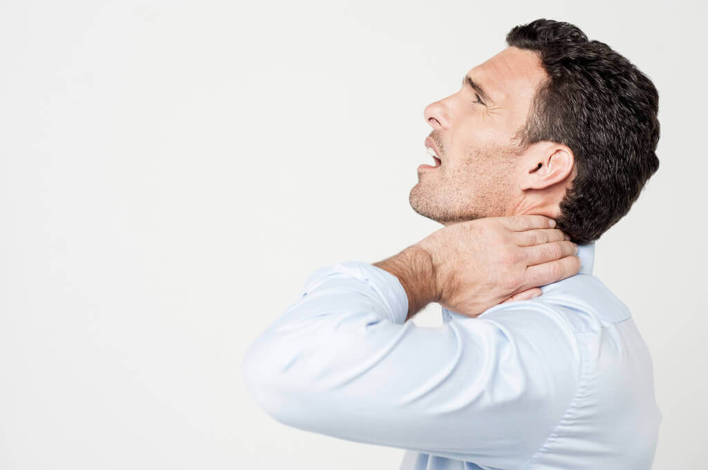
Torticollis means ‘twisted neck’. The neck becomes twisted to one side. The most common cause of torticollis is acute torticollis, also known as ‘wry neck’. Most of this leaflet is about the common acute torticollis. Other less common causes of torticollis are mentioned briefly later in the leaflet.
‘Acute’ means that the symptoms have developed quickly, over a period of hours, or often overnight. The twisting of the neck (torticollis) occurs when the muscles supporting the neck on one side are painful.
The cause of acute torticollis is often not known. It can happen in people with no previous neck symptoms. It is a common cause of neck pain in young people. There is usually no obvious injury.
However, it may be due to a minor sprain or irritation of a muscle or ligament in the neck. Some reasons for this include:
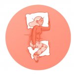
Sitting or sleeping in an unusual position without adequate neck support
Poor posture when looking at a computer screen.
Carrying heavy unbalanced loads (for example, a briefcase or shopping bag).
Allowing certain muscles of the neck to be exposed to cold (‘sleeping in a draught’).
It is common for people to go to bed feeling fine and to wake up the next morning with an acute torticollis.
The pain is usually on one side of the neck and stiffness of the muscles in that area twists the neck to one side. Attempts to straighten the neck are difficult due to pain. Occasionally, the pain is in the middle of the neck.
The pain may spread to the back of the head or the shoulder. The muscles of the affected side may be tender. Pressure on certain areas may trigger a ‘spasm’ of the muscle. Movement of the neck is restricted, particularly on one side
Diseases
-
Neck Pain
-
Shoulder & Elbow
-
Hand & Wrist
-
Arthritis
-
Lower Back Pain
-
Hip & Femur
-
Knee & Lower Leg
-
Foot & Ankle
-
Osteoporosis


