Hand & Wrist
Hand Anatomy
Few structures of the human anatomy are as unique as the hand. The hand needs to be mobile in order to position the fingers and thumb. Adequate strength forms the basis for normal hand function. The hand also must be coordinated to perform fine motor tasks with precision. The structures that form and move the hand require proper alignment and control in order for normal hand function to occur.
Important Structures
The important structures of the hand can be divided into several categories. These include
- bones and joints
- ligaments and tendons
- muscles
- nerves
- blood vessels
The front, or palm-side, of the hand is referred to as the palmar side. The back of the hand is called the dorsal side.
Bones and Joints
There are 27 bones within the wrist and hand. The wrist itself contains eight small bones, called carpals. The carpals join with the two forearm bones, the radius and ulna, forming the wrist joint. Further into the palm, the carpals connect to the metacarpals. There are five metacarpals forming the palm of the hand. One metacarpal connects to each finger and thumb. Small bone shafts called phalanges line up to form each finger and thumb.
The main knuckle joints are formed by the connections of the phalanges to the metacarpals. These joints are called the metacarpophalangeal joints (MCP joints). The MCP joints work like a hinge when you bend and straighten your fingers and thumb.
The three phalanges in each finger are separated by two joints, called interphalangeal joints (IP joints). The one closest to the MCP joint (knuckle) is called the proximal IP joint (PIP joint). The joint near the end of the finger is called the distal IP joint (DIP joint). The thumb only has one IP joint between the two thumb phalanges. The IP joints of the digits also work like hinges when you bend and straighten your fingers and thumb.
The joints of the hand, fingers, and thumb are covered on the ends with articular cartilage. This white, shiny material has a rubbery consistency. The function of articular cartilage is to absorb shock and provide an extremely smooth surface to facilitate motion. There is articular cartilage essentially everywhere that two bony surfaces move against one another, or articulate.
Ligaments and Tendons
Ligaments are tough bands of tissue that connect bones together. Two important structures, called collateral ligaments, are found on either side of each finger and thumb joint. The function of the collateral ligaments is to prevent abnormal sideways bending of each joint.
In the PIP joint (the middle joint between the main knuckle and the DIP joint), the strongest ligament is the volar plate. This ligament connects the proximal phalanx to the middle phalanx on the palm side of the joint. The ligament tightens as the joint is straightened and keeps the PIP joint from bending back too far (hyperextending). Finger deformities can occur when the volar plate loosens from disease or injury.
The tendons that allow each finger joint to straighten are called the extensor tendons. The extensor tendons of the fingers begin as muscles that arise from the backside of the forearm bones. These muscles travel towards the hand, where they eventually connect to the extensor tendons before crossing over the back of the wrist joint. As they travel into the fingers, the extensor tendons become the extensor hood. The extensor hood flattens out to cover the top of the finger and sends out branches on each side that connect to the bones in the middle and end of the finger.
The place where the extensor tendon attaches to the middle phalanx is called the central slip. When the extensor muscles contract, they tug on the extensor tendon and straighten the finger. Problems occur when the central slip is damaged, as can happen with a tear.
Muscles
Many of the muscles that control the hand start at the elbow or forearm. They run down the forearm and cross the wrist and hand. Some control only the bending or straightening of the wrist. Others influence motion of the fingers or thumb. Many of these muscles help position and hold the wrist and hand while the thumb and fingers grip or perform fine motor actions.
Most of the small muscles that work the thumb and pinky finger start on the carpal bones. These muscles connect in ways that allow the hand to grip and hold. Two muscles allow the thumb to move across the palm of the hand, an important function called thumb opposition.
The smallest muscles that originate in the wrist and hand are called the intrinsic muscles. The intrinsic muscles guide the fine motions of the fingers by getting the fingers positioned and holding them steady during hand activities.
Nerves
All of the nerves that travel to the hand and fingers begin together at the shoulder: the radial nerve, the median nerve, and the ulnar nerve. These nerves carry signals from the brain to the muscles that move the arm, hand, fingers, and thumb. The nerves also carry signals back to the brain about sensations such as touch, pain, and temperature.
The radial nerve runs along the thumb-side edge of the forearm. It wraps around the end of the radius bone toward the back of the hand. It gives sensation to the back of the hand from the thumb to the third finger. It also supplies the back of the thumb and just beyond the main knuckle of the back surface of the ring and middle fingers.
The median nerve travels through a tunnel within the wrist called the carpal tunnel. This nerve gives sensation to the thumb, index finger, long finger, and half of the ring finger. It also sends a nerve branch to control the thenar muscles of the thumb. The thenar muscles help move the thumb and let you touch the pad of the thumb to the tips each of each finger on the same hand, a motion called opposition.
The ulnar nerve travels through a separate tunnel, called Guyon’s canal. This tunnel is formed by two carpal bones, the pisiform and hamate, and the ligament that connects them. After passing through the canal, the ulnar nerve branches out to supply feeling to the little finger and half the ring finger. Branches of this nerve also supply the small muscles in the palm and the muscle that pulls the thumb toward the palm.
The nerves that travel to the hand are subject to problems. Constant bending and straightening of the wrist and fingers can lead to irritation or pressure on the nerves within their tunnels and cause problems such as pain, numbness, and weakness in the hand, fingers, and thumb.
Blood Vessels
Traveling along with the nerves are the large vessels that supply the hand with blood. The largest artery is the radial artery that travels across the front of the wrist, closest to the thumb. The radial artery is where the pulse is taken in the wrist. The ulnar artery runs next to the ulnar nerve through Guyon’s canal (mentioned earlier). The ulnar and radial arteries arch together within the palm of the hand, supplying the front of the hand, fingers, and thumb. Other arteries travel across the back of the wrist to supply the back of the hand, fingers, and thumb.
Wrist Anatomy
The anatomy of the wrist joint is extremely complex, probably the most complex of all the joints in the body. The wrist is actually a collection of many bones and joints. These bones and joints let us use our hands in lots of different ways. The wrist must be extremely mobile to give our hands a full range of motion. At the same time, the wrist must provide the strength for heavy gripping.
Important Structures
The important structures of the wrist can be divided into several categories. These include
- bones and joints
- ligaments and tendons
- muscles
- nerves
- blood vessels
Bones and Joints
There are 15 bones that form connections from the end of the forearm to the hand. The wrist itself contains eight small bones, called carpal bones. These bones are grouped in two rows across the wrist. The proximal row is where the wrist creases when you bend it. Beginning with the thumb-side of the wrist, the proximal row of carpal bones is made up of the scaphoid, lunate, and triquetrum. The second row of carpal bones, called the distal row, meets the proximal row a little further toward the fingers. The distal row is made up of the trapezium, trapezoid, capitate, hamate, and pisiform bones.
The proximal row of carpal bones connects the two bones of the forearm, the radius and the ulna, to the bones of the hand. The bones of the hand are called the metacarpal bones. These are the long bones that lie within the palm of the hand. The metacarpals attach to the phalanges, which are the bones in the fingers and thumb.
One reason that the wrist is so complicated is because every small carpal bone forms a joint with the bone next to it. This means that what we call the wrist joint is actually made up of many small joints.
Articular cartilage is the material that covers the ends of the bones of any joint. Articular cartilage can be up to one-quarter of an inch thick in the large, weight-bearing joints. It is thinner in joints such as the wrist that don’t support a lot of weight. Articular cartilage is white, shiny, and has a rubbery consistency. It is slippery, which allows the joint surfaces to slide against one another without causing any damage.
The function of articular cartilage is to absorb shock and provide an extremely smooth surface to make motion easier. We have articular cartilage essentially everywhere that two bony surfaces move against one another, or articulate. In the wrist, articular cartilage covers the sides of all the carpals and the ends of the bones that connect from the forearm to the fingers.
Ligaments and Tendons
Ligaments are soft tissue structures that connect bones to bones. The ligaments around a joint usually combine to form a joint capsule. A joint capsule is a watertight sac that surrounds a joint and contains lubricating fluid called synovial fluid. In the wrist, the eight carpal bones are surrounded and supported by a joint capsule.
Two important ligaments support the sides of the wrist. These are the collateral ligaments. There are two collateral ligaments that connect the forearm to the wrist, one on each side of the wrist.
As its name suggests, the ulnar collateral ligament (UCL) is on the ulnar side of the wrist. It crosses the ulnar edge (the side away from the thumb) of the wrist. It starts at the ulnar styloid, the small bump on the edge of the wrist (on the side away from the thumb) where the ulna meets the wrist joint. There are two parts to the cord-shaped UCL. One part connects to the pisiform (one of the small carpal bones) and to the transverse carpal ligament, a thick band of tissue that crosses in front of the wrist. The other goes to the triquetrum (a small carpal bone near the ulnar side of the wrist). The UCL adds support to a small disc of cartilage where the ulna meets the wrist. This structure is called the triangular fibrocartilage complex (TFCC) and is discussed in more detail below. The UCL stabilizes the TFCC and keeps the wrist from bending too far to the side (toward the thumb).
The radial collateral ligament (RCL) is on the thumb side of the wrist. It starts on the outer edge of the radius on a small bump called the radial styloid. It connects to the side of the scaphoid, the carpal bone below the thumb. The RCL prevents the wrist from bending too far to the side (away from the thumb).
Just as there are many bones that form the wrist, there are many ligaments that connect to and support these bones. Injury or problems that cause these ligaments to stretch or tear can eventually lead to arthritis in the wrist.
At the wrist, the end of the ulna bone of the forearm articulates with two carpal bones, the lunate and the triquetrum. A unique structure mentioned earlier, the triangular fibrocartilage complex (TFCC), sits between the ulna and these two carpal bones. The TFCC is a small cartilage pad that cushions this part of the wrist joint. It also improves the range of motion and gliding action within the wrist joint.
There are several important tendons that cross the wrist. Tendons connect muscles to bone. The tendons that cross the wrist begin as muscles that start in the forearm. Those that cross the palm side of the wrist are the flexor tendons. They curl the fingers and thumb, and they bend the wrist. The flexor tendons run beneath the transverse carpal ligament (mentioned earlier). This structure lies on the palm side of the wrist. This band of tissue keeps the flexor tendons from bowing outward when you curl your fingers, thumb, or wrist. The tendons that travel over the back of the wrist, the extensor tendons, run through a series of tunnels, called compartments. These compartments are lined with a slick substance called tenosynovium, which prevents friction as the extensor tendons glide inside their compartment.
Muscles
The main muscles that are important at the wrist have been mentioned above in the discussion about tendons. These muscles generally start further up in the forearm. The tendons of these muscles cross the wrist. They control the actions of the fingers, thumb, and wrist.
Nerves
All of the nerves that travel to the hand cross the wrist. Three main nerves begin together at the shoulder: the radial nerve, the median nerve, and the ulnar nerve. These nerves carry signals from the brain to the muscles that move the arm, hand, fingers, and thumb. The nerves also carry signals back to the brain about sensations such as touch, pain, and temperature.
The radial nerve runs along the thumb-side edge of the forearm. It wraps around the end of the radius bone toward the back of the hand. It gives sensation to the back of the hand from the thumb to the third finger. It also goes to the back of the thumb and just beyond the main knuckle of the back surface of the ring and middle fingers.
The median nerve travels through a tunnel within the wrist called the carpal tunnel. The median nerve gives sensation to the palm sides of the thumb, index finger, long finger, and half of the ring finger. It also sends a nerve branch to control the thenar muscles of the thumb. The thenar muscles help move the thumb and let you touch the pad of the thumb to the tips each of each finger on the same hand, a motion called opposition.
The ulnar nerve travels through a separate tunnel, called Guyon’s canal. This tunnel is formed by two carpal bones (the pisiform and hamate), and the ligament that connects them. After passing through the canal, the ulnar nerve branches out to supply feeling to the little finger and half the ring finger. Branches of this nerve also supply the small muscles in the palm and the muscle that pulls the thumb toward the palm.
The nerves that travel through the wrist are subject to problems. Constant bending and straightening of the wrist and fingers can lead to irritation or pressure on the nerves within their tunnels and cause problems such as pain, numbness, and weakness in the hand, fingers, and thumb.
Blood Vessels
Traveling along with the nerves are the large vessels that supply the hand with blood. The largest artery is the radial artery that travels across the front of the wrist, closest to the thumb. The radial artery is where the pulse is taken in the wrist. The ulnar artery runs next to the ulnar nerve through Guyon’s canal (mentioned earlier). The ulnar and radial arteries arch together within the palm of the hand, supplying the front of the hand and fingers. Other arteries travel across the back of the wrist to supply the back of the hand and fingers.
Introduction
Carpal tunnel syndrome (CTS) is a common problem affecting the hand and wrist. Symptoms begin when the median nerve gets squeezed inside the carpal tunnel of the wrist, a medical condition known as nerve entrapment or compressive neuropathy. Any condition that decreases the size of the carpal tunnel or enlarges the tissues inside the tunnel can produce the symptoms of CTS.
This syndrome has received a lot of attention in recent years because of suggestions that it may be linked with occupations that require repeated use of the hands, such as typing on a computer keyboard or doing assembly work. Actually, many people develop this condition regardless of the type of work they do.
Anatomy
Where is the carpal tunnel, and what does it do?
The carpal tunnel is an opening through the wrist to the hand that is formed by the bones of the wrist on one side and the transverse carpal ligament on the other. (Ligaments connect bones together.) This opening forms the carpal tunnel.
The median nerve passes through the carpal tunnel into the hand. It gives sensation to the thumb, index finger, long finger, and half of the ring finger. It also sends a nerve branch to control the thenar muscles of the thumb. The thenar muscles help move the thumb and let you touch the pad of the thumb to the tips of each finger on the same hand, a motion called opposition.
The median nerve and flexor tendons pass through the carpal tunnel. The median nerve rests on top of the tendons, just below the transverse carpal ligament. The flexor tendons are important because they allow movement of the fingers, thumb, and hand, such as when grasping. The tendons are covered by a material called tenosynovium. The tenosynovium is a slippery covering that allows the tendons to glide next to each other as they contract and relax to move the hand and fingers.
Causes
What causes CTS?
Any condition that makes the area inside the carpal tunnel smaller or increases the size of the tissues within the tunnel can lead to symptoms of CTS. The carpal tunnel cannot expand so any condition that causes abnormal pressure in the tunnel can produce symptoms of CTS. And any increase in pressure within the carpal tunnel can reduce blood flow to the nerve, leading to loss of nerve function.
Various types of arthritis can cause swelling and pressure in the carpal tunnel. The way people do their tasks can put them at risk for problems of CTS. Some of these risks include
- force
- posture
- wrist alignment
- repetition
- temperature
- vibration
One of these risks alone may not cause a problem. But doing a task that involves several factors may pose a greater risk. And the longer a person is exposed to one or more risks, the greater the possibility of having a problem with CTS. However, scientists believe that other factors such as smoking, obesity, and caffeine intake may actually be more important in determining whether a person is more likely to develop CTS.
In other instances, CTS can start when the tenosynovium thickens from irritation or inflammation. This thickening causes pressure to build inside the carpal tunnel. But the tunnel can’t stretch any larger in response to the added swelling, so the median nerve starts to squeeze against the transverse carpal ligament. If the pressure continues to build up, the nerve is eventually unable to function normally.
When pressure builds on the median nerve, the blood supply to the outer covering of the nerve slows down and may even be cut off. The medical term for this is ischemia. At first, only the outside covering of the nerve is affected. But if the pressure keeps building up, the inside of the nerve will start to become thickened. New cells (called fibroblasts) form within the nerve and create scar tissue. This is thought to produce the feelings of pain and numbness in the hand. If pressure is taken off right away, the symptoms will go away quickly. Pressure that isn’t eased right away can slow or even stop the chances for recovery.
Trauma such as a wrist fracture, fracture/dislocation, infection, burns or other thermal injuries, bleeding disorders, and high-pressure injection injuries can result in a condition called i>acute carpal tunnel syndrome. This is much less common than the chronic compressive neuropathy caused by any of the risk factors described in this document.
A traumatic wrist injury may cause swelling and extra pressure within the carpal tunnel. The area inside the tunnel can also be reduced after a wrist fracture or dislocation if the bone pushes into the tunnel. Fractured wrist bones may later cause CTS if the healed fragments result in abnormal irritation on the flexor tendons.
Other conditions in the body can produce symptoms of CTS. Pregnancy can cause fluid to be retained, leading to extra pressure in the carpal tunnel. Diabetics may report symptoms of CTS, which may be from a problem in the nerve (called neuropathy) or from actual pressure on the median nerve. People with low thyroid function (called hypothyroidism) are more prone to problems of CTS. Tumors or cysts in the wrist, on the tendons, or in the carpal tunnel can also cause CTS.
Symptoms
What does CTS feel like?
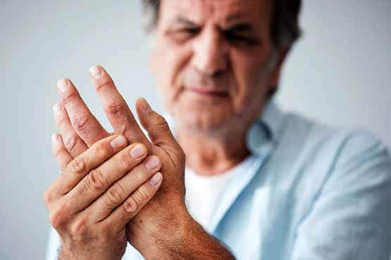
One of the first symptoms of CTS is gradual tingling and numbness in the areas supplied by the median nerve. This is typically followed by dull, vague pain where the nerve gives sensation in the hand. The hand may begin to feel like it’s asleep, especially in the early morning hours after a night’s rest.
In the case of acute CTS, symptoms are sudden and severe, occurring over a matter of hours rather than weeks or months with the more chronic form of this condition.
Whether acute or chronic, pain associated with carpal tunnel syndrome may even spread up the arm to the shoulder. If the condition progresses, the thenar muscles of the thumb can weaken, causing the hand to be clumsy when picking up a glass or cup. If the pressure keeps building in the carpal tunnel, the thenar muscles may begin to atrophy (shrink).
Touching the pad of the thumb to the tips of the other fingers becomes difficult, making it hard to grasp items such as a steering wheel, newspaper, or telephone.
Diagnosis
How do doctors identify the condition?
Your doctor begins the evaluation by obtaining a history of the problem, followed by a thorough physical examination. Your description of the symptoms and the physical examination are the most important parts in the diagnosis of CTS. Commonly, patients will complain first of waking in the middle of the night with pain and a feeling that the whole hand is asleep.
Careful investigation usually shows that the little finger is unaffected. This can be a key piece of information to make the diagnosis. If you awaken with your hand asleep, pinch your little finger to see if it is numb also, and be sure to tell your doctor if it is or isn’t. Other complaints include numbness while using the hand for gripping activities, such as sweeping, hammering, or driving.
If your symptoms started after a traumatic wrist injury, X-rays may be needed to check for a fractured bone or a fracture with dislocation.
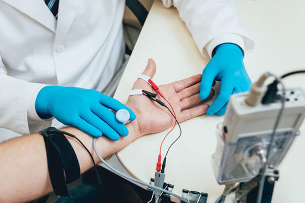
If more information is needed to make the diagnosis, electrical studies of the nerves in the wrist may be requested by your doctor. Several tests are available to see how well the median nerve is functioning, including the nerve conduction velocity (NCV) test. This test measures how fast nerve impulses move through the nerve
Introduction
Trigger finger and trigger thumb are conditions affecting the movement of the tendons as they bend the fingers or thumb toward the palm of the hand. This movement is called flexion.
Anatomy
Where does the condition develop?
The tendons that move the fingers are held in place on the bones by a series of ligaments called pulleys. These ligaments form an arch on the surface of the bone that creates a sort of tunnel for the tendon to run in along the bone. To keep the tendons moving smoothly under the ligaments, the tendons are wrapped in a slippery coating called tenosynovium. The tenosynovium reduces the friction and allows the flexor tendons to glide through the tunnel formed by the pulleys as the hand is used to grasp objects.
Causes
Why do I have this problem?
Triggering is usually the result of a thickening in the tendon that forms a nodule, or knob. The pulley ligament may thicken as well. The constant irritation from the tendon repeatedly sliding through the pulley causes the tendon to swell in this area and create the nodule. Rheumatoid arthritis, partial tendon lacerations, repeated trauma from pistol-gripped power tools, or long hours grasping a steering wheel can cause triggering. Infection or damage to the synovium causes a rounded swelling (nodule) to form in the tendon.
Triggering can also be caused by a congenital defect that forms a nodule in the tendon. The condition is not usually noticeable until infants begin to use their hands.
Symptoms
What does a trigger finger or thumb feel like?
The symptoms of trigger finger or thumb include pain and a funny clicking sensation when the finger or thumb is bent. Pain usually occurs when the finger or thumb is bent and straightened. Tenderness usually occurs over the area of the nodule, at the bottom of the finger or thumb. The clicking sensation occurs when the nodule moves through the tunnel formed by the pulley ligaments. With the finger straight, the nodule is at the far edge of the surrounding ligament. When the finger is flexed, the nodule passes under the ligament and causes the clicking sensation. If the nodule becomes too large it may pass under the ligament, but it gets stuck at the near edge. The nodule cannot move back through the tunnel, and the finger is locked in the flexed trigger position.
Diagnosis
How do doctors identify the condition?
The diagnosis of trigger finger and thumb is usually quite obvious on physical examination. Usually a palpable click can be felt as the nodule snaps under the first finger pulley. If the condition is allowed to progress, the nodule may swell to the point where it gets caught and the finger is locked in a bent, or flexed, position. No special tests or X-rays are required.
A ganglion is a small, harmless cyst, or sac of fluid, that sometimes develops in the wrist. Doctors don’t know exactly what causes ganglions, but a ganglion that isn’t painful and doesn’t interfere with activity can often be left untreated without harm to the patient. However, treatment options are available for painful ganglions or ones that cause problems.
Anatomy
What parts of the wrist are involved?
The anatomy of the wrist joint is extremely complex, probably the most complex of all the joints in the body. The wrist is actually a collection of many joints and bones. These joints and bones let us use our hands in lots of different ways. The wrist must be extremely mobile to give our hands a full range of motion. At the same time, the wrist must provide the strength for heavy gripping.
The wrist is made of eight separate small bones, called the carpal bones. The carpal bones connect the two bones of the forearm, the radius and the ulna, to the bones of the hand. The metacarpal bones are the long bones that lie underneath the palm.
Ligaments connect and hold all these wrist bones together. The ligaments allow the bones to move in all directions. These ligaments meld together to form the joint capsule of the wrist. The joint capsule is a watertight sac of tissue that surrounds the wrist bones. Inside the wrist capsule are the joints themselves. The joint capsule contains a small amount of lubricant, called synovial fluid, that allows the bones to move together easily. The many tendons required to move the fingers run just outside the joint capsule.
Ganglions are generally attached by a stalk of tissue to a nearby joint capsule, tendon, or tendon sheath (tissue covering the tendon). Wrist ganglions are attached to the wrist joint capsule. Typically only one ganglion appears, often in a location that is predictable to doctors. However, ganglions have been seen in almost every joint in the hand and wrist.
Sixty to 70 percent of wrist ganglions are dorsal wrist ganglions. A dorsal wrist ganglion is found on the back of the hand, often centered over the wrist, though it can appear in any number of areas along the back of the wrist. A dorsal wrist ganglion may be not be visible from the outside. Doctors refer to this hidden type of ganglion as occult, or concealed.
A volar wrist ganglion typically appears on the palm side of the wrist in the wrist crease just below the thumb. This is the second most common type of wrist ganglion.
Causes
Why do I have this problem?
Doctors don’t know why ganglions develop. In some cases, the wrist has been injured previously. Repetitive injuries, such as those that can occur from playing tennis or golf frequently, seem to play a role in ganglion development as well.
One theory suggests that wrist ganglions are formed when connective tissue degenerates or is damaged by wear and tear. The damaged tissue forms a weakened spot in the joint capsule, just like a weak spot on a car tire that allows the inner tube to bulge through. The joint fluid may escape through this weakened area and begin to collect in a cyst outside the joint. Over time this cyst grows larger. The joint fluid seems to move out of the wrist joint into the ganglion, but not the other way. In the end, a clear, sticky fluid fills the cyst. The fluid is a mix of chemicals normally found in the joint.
Symptoms
What does a ganglion feel like?
A patient with a dorsal wrist ganglion may feel a bump or mass on the back of the wrist. With a volar wrist ganglion, the bump is felt on the wrist crease below the thumb. The mass may appear suddenly, or it may develop over time. The ganglion may occasionally increase or decrease in size.
The wrist may ache or feel tender. The ganglion may also interfere with activities. A volar wrist ganglion may compress the median or ulnar nerve, causing trouble with sensation and movement. An occult dorsal wrist ganglion may be quite painful and tender, even though it is smaller than other ganglions. Typically the symptoms from a ganglion are not harmful and generally do not grow worse. These cysts will not turn into cancer.
Diagnosis
Your doctor will ask for a history of the problem and examine your hand and wrist. Usually, this is all that’s required to diagnose a ganglion. An occult dorsal wrist ganglion, however, may be more difficult to locate because of its small size.
Intersection syndrome is a painful condition of the forearm and wrist. It can affect people who do repeated wrist actions, such as weight lifters, downhill skiers, and canoeists. Heavy raking or shoveling can also cause intersection syndrome.
Anatomy
What part of the forearm is causing my pain?
The pain from intersection syndrome is usually felt on the top of the forearm, about three inches above the wrist. At this spot, two muscles that connect to the thumb cross over (or intersect) the two underlying wrist tendons (tendons connect muscles to bones).
The two muscles that cross over the wrist tendons control the thumb. They are the extensor pollicis brevis and the abductor pollicis longus. These two muscles start on the forearm, cross over the two wrist tendons, and connect on the back part of the thumb. When these muscles work, they pull the thumb out and back.
The extensor carpi radialis brevis and the extensor carpi radialis longus muscles run lengthwise along the back of the forearm. The tendons of these two muscles attach on the back of the hand. The action of these two wrist tendons pulls the wrist back, into extension.
Most of the tendons around the wrist are covered with a thin tissue called tenosynovium. Tenosynovium is very slippery. It allows tendons to glide against one another and the surrounding muscles, fat, and skin with very little friction.
Causes
What caused my condition?
If you overuse the wrist extensor tendons, the slippery tenosynovial lining may become inflamed from the constant rubbing against the two thumb muscles. As the tenosynovium becomes more irritated and inflamed, it swells and thickens. You feel pain when you move your wrist because the swollen tendons are rubbing against the thumb muscles.
Wrist extensor tendons work like the bow used by violin players. The wrist extensor tendons are like the bow, and the thumb muscles are like the strings. As the wrist curls down and in, the wrist tendons rub back and forth against the thumb muscles. The friction builds up, much like the effect of rubbing two sticks together. This leads to irritation and inflammation of the tenosynovium covering the wrist extensor tendons.
The wrist extensor tendons are strained by any activities that cause the wrist to curl down and in, toward the thumb. These wrist movements are especially common in downhill skiers when they plant their ski poles deeply in powder snow. The same movement is involved when pulling a rake against hard ground. Racket sports, weight lifting, canoeing, and rowing can also stress the wrist extensor tendons.
Symptoms
What does intersection syndrome feel like?
The friction on the wrist tendons causes pain and swelling in the tenosynovium that covers the tendons. The friction hampers the smooth gliding action. You may hear a squeaking sound and feel creaking as the tendons rub against the muscles. This is called crepitus. You may have swelling and redness at the intersection point. Pain can spread down to the thumb or up along the edge of the forearm.
Diagnosis
What tests will my doctor run?
Doctors usually make the diagnosis of intersection syndrome during a physical examination. Most of the time no special tests are required.
The main challenge is distinguishing intersection syndrome from de Quervain’s tenosynovitis. De Quervain’s tenosynovitis is a condition that is very similar to intersection syndrome.
Both syndromes involve inflammation in the tendons of the wrist. However, the pain begins in different spots. Intersection syndrome causes pain at the intersection point, about three inches up the forearm. De Quervain’s tenosynovitis causes pain along the edge of the wrist, closer to the hand. Your doctor will examine your forearm and wrist carefully to locate exactly where your pain is coming from.
A sprained wrist is a relatively common condition characterized by damage or tearing of the connective tissue (such as ligaments, cartilage and joint capsule) of the wrist.
The wrist comprises the joining of 8 small bones in the hand and the two bones of the forearm (radius and ulna) (figure 1). Between the wrist bones lie many small joints, each of which comprises of strong connective tissue wrapping around the bony ends and cartilage which lies between the joint surfaces, cushioning the impact of one bone on another during activity.
During certain movements of the wrist, stretching or compression forces are placed on the joints of the wrist. If these forces are excessive due to too much repetition or high force, injury to the joints may occur. This may involve damage to the cartilage or tearing to the connective tissue surrounding the joint. When this occurs, the condition is known as a sprained wrist.
Causes of a sprained wrist
A sprained wrist most commonly occurs due to a specific incident such as a fall onto an outstretched hand. This may occur with any fall, but is particularly common in sports such as cycling, skateboarding or snowboarding (particularly in icy conditions) where a fall onto a hard surface is unforgiving. A wrist sprain may also occur during weight lifting or boxing particularly in those with poor lifting or punching technique. Occasionally, a sprained wrist may occur due to repetitive strain associated with overuse. This may be the case in patients who perform heavy manual work involving the wrist such as the repetitive use of a hammer or screw driver.
Signs and symptoms of a sprained wrist
Patients with a sprained wrist often experience a sudden onset of wrist pain during the causative activity. However, patients may also experience pain and stiffness after the provocative activity, particularly the next morning. Symptoms may be felt on the front, back or sides of the wrist. Occasionally pain may be referred into the forearm or hand on the affected side. Symptoms are generally exacerbated with heavy activities that involve use of the hand and wrist such as opening jars or doors, picking up heavy objects, general gripping activity or placing weight through the affected hand (e.g. doing push-ups). It is also common for patients to experience pain on firmly touching the affected region, and, in some cases, a feeling of weakness in the wrist and hand may also be present.
Diagnosis of a sprained wrist
A thorough subjective and objective examination from a physiotherapist is usually sufficient to diagnose a sprained wrist. Investigations such as an X-ray, MRI or CT scan may be required to assist diagnosis and rule out other injuries (particularly fractures).
Kienbock’s disease is a condition in which one of the small bones of the wrist loses its blood supply and dies, causing pain and stiffness with wrist motion. In the late stages of the disease, the bone collapses, shifting the position of other bones in the wrist. This shifting eventually leads to degenerative changes and osteoarthritis in the joint. While the exact cause of this uncommon disease isn’t known, a number of treatment options are available.
Anatomy
How does the wrist joint work?
The anatomy of the wrist joint is extremely complex, probably the most complex of all the joints in the body. The wrist is actually a collection of many joints and bones. These joints and bones let us use our hands in lots of different ways. The wrist must be extremely mobile to give our hands a full range of motion. At the same time, the wrist must provide the strength for heavy gripping.
The wrist is made of eight separate small bones, called the carpal bones. The lunate is one of these bones.
It is the bone that is affected in patients with Kienbock’s disease.
The carpal bones connect the two bones of the forearm, the radius and the ulna, to the bones of the hand. The metacarpal bones are the long bones that lie underneath the palm. The metacarpals attach to the phalanges, which are the bones in the fingers and thumb.
Causes
Why do I have this condition?
Doctors have not determined exactly what causes Kienbock’s disease. A number of factors seem to be involved. Usually the patient has injured the wrist. The injury may be a single incident, such as a sprain, or a repetitive trauma. But the injury alone does not seem to cause the disease.
The way that blood vessels supply the lunate is thought to play a role in Kienbock’s disease. Some bones in the body simply have fewer blood vessels that bring in blood. The lunate is one of those bones. A bone with a limited blood supply may be more at risk of developing the disease after an injury. The reduced blood supply might be the result of a previous injury to the blood vessels.
Other bones around the lunate may play a role in the disease, too. The length of the ulna, the bone of the forearm on the opposite side of the thumb, may be a factor. When the ulna is shorter than the radius, an imbalance of pressure is created in the wrist joint. Normally, the ulna supports a portion of the force that needs to be transferred from the hand to the forearm. If the ulna is too short, this cannot occur. The lunate is caught between the capitate bone and the radius and must absorb more force when the hand is used for heavy gripping activities. Over time, this extra force may make it more likely for a person to develop Kienbock’s disease. Chronic repetitive trauma can lead to damage of the arteries supplying blood to the lunate.
Kienbock’s disease is also sometimes found in people with other medical conditions that are known to damage small blood vessels of the body. Whatever the cause, the lunate bone develops a condition called osteonecrosis. In osteonecrosis, the bone dies, usually because it’s not getting enough blood.
Symptoms
What does Kienbock’s disease feel like?
The primary symptoms of Kienbock’s disease are pain in the wrist and limited wrist motion. Pain may vary from slight discomfort to constant pain. In the early stages there may be pain only during or after heavy activity using the wrist. The pain usually gets slowly worse over many years. The wrist may swell. The area over the back of the wrist near the lunate bone may feel tender. You may not be able to move your wrist as much as normal or grip objects as well.
Patients often have the condition for months or years before seeking treatment. It rarely affects both wrists. Without treatment the bone may collapse. When the lunate bone is displaced or fragmented, it can rub on the tendons that slide along the back of the wrist – the extensor tendons. The abnormal bone may eventually wear through one or more of the extensor tendons along the back of the wrist. The wrist becomes unstable. The resulting misalignment causes even more uneven wear on the bones leading to osteoarthritis between the radius and the carpal bones.
Diagnosis
How do doctors identify the problem?
Your doctor will begin by taking a detailed history of the problem and examining the wrist.
X-rays and possibly a magnetic resonance imaging (MRI) scan will be ordered. The X-rays are useful to determine how far the disease has advanced. This helps your doctor plan treatment. The MRI machine uses magnetic waves instead of radiation to take a series of pictures that look like slices of the wrist. The MRI scan is most useful if your doctor is not sure whether the lunate bone has lost its blood supply. The MRI is extremely accurate at showing whether a bone has a blood supply or not. Changes in the lunate bone will usually appear on one of these tests. No other tests are usually required.
Guyon’s canal syndrome is an entrapment of the ulnar nerve as it passes through a tunnel in the wrist called Guyon’s canal. This problem is similar to carpal tunnel syndrome but involves a completely different nerve. Sometimes both conditions can cause a problem in the same hand.
Anatomy
Where is the ulnar nerve, and what does it do?
The ulnar nerve actually starts at the side of the neck, where the individual nerve roots exit the spine through small openings between the vertebrae. The nerve roots then join together to form three main nerves that travel down the arm to the hand, one of which is the ulnar nerve.
After leaving the side of the neck, the ulnar nerve travels through the armpit and down the arm to the hand and fingers. As it crosses the wrist, the ulnar nerve and ulnar artery run through the tunnel known as Guyon’s canal. This tunnel is formed by two bones (the pisiform and hamate) and the ligament that connects them. After passing through the canal, the ulnar nerve branches out to supply feeling to the little finger and half the ring finger. Branches of this nerve also supply the small muscles in the palm and the muscle that pulls the thumb toward the palm.
The hamate bone forms one side of Guyon’s canal. This bone has a small hook-shaped spur that sticks out to provide an attachment for several wrist ligaments. Known as the hook of hamate, this small bone can break off and press against the ulnar nerve within Guyon’s canal.
Causes
Why do I have this problem?
Guyon’s canal syndrome has several causes. Overuse of the wrist from heavy gripping, twisting, and repeated wrist and hand motions can cause symptoms. Working with the hand bent down and outward can squeeze the nerve inside Guyon’s canal.
Constant pressure on the palm of the hand can produce symptoms. This is common in cyclists and weight lifters from the pressure of gripping. It can also happen after running a jackhammer or when using crutches.
Pressure or irritation of the ulnar nerve can cause symptoms of Guyon’s canal syndrome. A traumatic wrist injury may cause swelling and extra pressure on the ulnar nerve within the canal. Arthritis in the wrist bones and joints may eventually irritate and compress the ulnar nerve. In rare cases, the ulnar artery that travels right beside the nerve may be damaged and form a blood clot. The symptoms caused by the clot mimic Guyon’s canal syndrome. The lack of blood supply to the ulnar nerve is believed to cause the symptoms.
As mentioned earlier, a fractured hamate bone in the wrist can pinch the nerve inside Guyon’s canal. This bone is sometimes fractured when golfers club the ground instead of the golf ball and when baseball players are batting.
Symptoms
What does Guyon’s canal syndrome feel like?
Symptoms usually begin with a feeling of pins and needles in the ring and little fingers, which is often noticed in the early morning when first awakening. This may progress to a burning pain in the wrist and hand followed by decreased sensation in the ring and little fingers. The hand may become clumsy when the muscles controlled by the ulnar nerve become weak. Weakness can affect the small muscles in the palm of the hand and the muscle that pulls the thumb into the palm. Gradual weakness in these muscles makes it hard to spread your fingers and pinch with your thumb.
This syndrome is much less common than carpal tunnel syndrome (CTS), yet both conditions can occur at the same time. The numbness caused by these two syndromes affects the hand in different locations. When the median nerve is compressed in CTS, pain and numbness spread into the thumb, index finger, middle finger, and half of the ring finger. Compression of the ulnar nerve in Guyon’s canal syndrome usually causes numbness in the pinky and half of the ring finger.
Diagnosis
How do doctors identify the problem?
The diagnosis of Guyon’s canal syndrome begins with a careful history and physical examination by your doctor. Compression can occur at several areas along the ulnar nerve, and your doctor will test to find exactly where the nerve is being affected. If it is unclear on the physical examination where the nerve is being squeezed, electrical studies may be ordered to try to find the area of compression.
Nerve conduction velocity (NCV) is a test that measures how fast nerve impulses travel along the nerve. Your doctor might want this test to be done to help pinpoint your problem. Special tests may be required to study the nerve.
The NCV is sometimes combined with an electromyogram (EMG). The EMG is done by testing the muscles of the forearm that are controlled by the ulnar nerve to determine if they are working properly. If the test shows a problem with the muscle, the nerve that goes to the muscle might not be working correctly. This is similar to checking whether the wiring in a lamp is working. If the light still doesn’t work after you’ve put in a new bulb, you can begin to tell if there’s a problem in the wiring.
If your symptoms started after a traumatic wrist injury, X-rays may be needed to check for a fractured or dislocated bone
Doctors commonly diagnose a sprained wrist after a patient falls on an outstretched hand. However, if pain and swelling don’t go away, doctors become suspicious that the injury is actually more serious. A fall on an outstretched hand commonly breaks the scaphoid bone of the wrist. X-rays taken at the time of the injury may not clearly show the fracture. If the fracture is not recognized early, it may not heal properly. This can lead to problems later.
Anatomy
Where is the scaphoid bone of the wrist?
The anatomy of the wrist joint is extremely complex, probably the most complex of all the joints in the body. The joint is actually a collection of many joints and many bones. These joints and bones let us use our hands in many ways. The wrist must be extremely mobile to give our hands a full range of motion. At the same time, the wrist must provide the strength for heavy gripping.
The wrist is made up of eight separate small bones, called the carpal bones. The scaphoid bone is a carpal bone near the base of the thumb. The carpal bones connect the two bones of the forearm, the radius and the ulna, to the bones of the hand. The metacarpal bones are the long bones that lie underneath the palm. The metacarpals attach to the phalanges, which are the bones in the fingers and thumb.
One reason that the wrist is so complicated is because every small bone forms a joint with the bone next to it. This means that what we call the wrist joint is actually made up of many small joints. Ligaments connect all the small bones to each other, and to the radius, ulna, and metacarpal bones.
The scaphoid bone is a small carpal bone on the thumb side (radial side) of the wrist. It is the most commonly fractured carpal bone. This is probably because it actually crosses two rows of carpal bones, forming a hinge. A fall on the outstretched hand puts heavy stress on the scaphoid bone. This stress can cause either a small crack through the middle of the bone or a complete separation of the bone into two pieces. A separation is called a displaced fracture.
Causes
What causes a scaphoid fracture?
A scaphoid fracture is almost always caused by a fall on the outstretched hand. We commonly try to break a fall by putting our hands out for protection. Landing on an outstretched hand makes hand and wrist injuries, including a fracture of the scaphoid bone, fairly common.
When a scaphoid fracture is recognized on the first X-ray, treatment begins immediately. But patients often assume that the injury is just a sprain, and they wait for it to heal on its own. In some cases, the wrist gets better. In many cases the bone fails to heal. The scaphoid fracture then develops into what surgeons call a nonunion.
A nonunion can occur in two ways. In a simple nonunion, the two pieces of bone fail to heal together. The second type of nonunion is much more serious. The lower half of the fractured bone loses its blood supply and actually dies. This condition is called avascular necrosis (Avascular means no blood supply, and necrosis means dead.)
The scaphoid bone is at risk for avascular necrosis. Only one small artery enters the bone, at the end that is closest to the thumb. If the fracture tears the artery, the blood supply is lost. Avascular necrosis becomes easy to see on X-rays several months after the injury.
Symptoms
How will I know if I have a scaphoid fracture?
The symptoms of a fresh fracture of the scaphoid bone usually include pain in the wrist and tenderness in the area just below the thumb. You may also see swelling around the wrist. The swelling occurs because blood from the fractured bone fills the wrist joint. Thin people will see a bulging of the joint capsule. The joint capsule is the watertight sac that encloses the joint.
Symptoms of a nonunion of the scaphoid bone are more subtle. You may have pain when you use your wrist. However, the pain may be very minimal. It is fairly common for doctors to see a nonunion of the scaphoid bone on X-rays, but the patient can’t remember an injury. These people probably suffered a wrist injury years ago that they thought was a simple sprain. Still, the most common symptom of a nonunion is a gradual increase in pain. Over several years the nonunion can lead to degenerative arthritis in the wrist joint.
Diagnosis
What tests will my doctor run?
Your doctor will first take a medical history. You will be asked questions about your pain and about any injuries to your wrist. Your doctor will also do a physical exam. The prodding and moving may hurt your wrist a bit. But it is important that your doctor know exactly where your pain is coming from.
Doctors should assume that any patient who has fallen on an outstretched hand and has swelling or tenderness on the thumb side of the wrist has a scaphoid fracture. You should assume this until tests prove otherwise. X-rays taken immediately after the injury may not show a fracture. Still, most surgeons will put a cast on the wrist and get another X-ray in 10 days. This gives the edges of the fractured bone time to heal, and may prevent nonunion. By waiting 10 days, the fracture is easier to see on an X-ray.
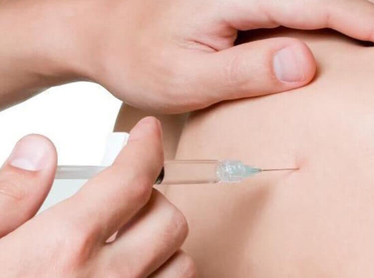
If it is still not clear whether or not you have a fracture, your doctor may order other imaging tests. You may have a bone scan done. A bone scan involves injecting tracers into your blood stream. The tracers then show up on special X-rays of your wrist. The tracers build up in areas of extra stress to bone tissue, such as a fracture.
Your doctor may also order a magnetic resonance imaging (MRI) scan. An MRI scan is a special imaging test that uses magnetic waves to create pictures of your body in slices. The MRI scan shows tendons as well as bones. It is painless and requires no needles or injections.
Ulnar nerve compression is a condition characterized by pressure being placed on the ulnar nerve as it passes along the inner aspect of the elbow (funny bone).
The ulna nerve originates from several spinal nerves in the neck and travels through the upper arm, into the forearm, hand and fingers. It passes the elbow in a shallow groove situated at the inner aspect of the elbow between the medial epicondyle and olecranon (figure 1). The ulna nerve is responsible for supplying sensation to the inner aspect of the forearm, the palm of the hand on the little finger side, the little finger and half of the ring finger. It is also responsible for supplying some muscles of the forearm and hand.
Following a direct impact to the inner aspect of the elbow, the ulnar nerve may be compressed. In addition, damage and inflammation of tissue adjacent to the ulnar nerve may also place pressure on the nerve. When this occurs the condition is known as ulnar nerve compression.
Ulnar nerve compression can occur at any age and is more common in the dominant arm especially in the case of overuse injuries. Occasionally, it may affect both arms.
Causes of ulnar nerve compression
Ulnar nerve compression most commonly occurs due to a direct impact to the nerve as it passes the inner aspect of the elbow. This is particularly common during a fall onto the elbow or due to a direct impact from an object or person. This may injure and compress the nerve directly or damage adjacent tissue causing inflammation, swelling and subsequent secondary compression of the nerve. Adjacent tissue may also be damaged due to overuse (such as excessive gripping activities) again placing pressure on the ulnar nerve due to inflammation, swelling and scar tissue formation.
Ulnar nerve compression may also occur due to excessive traction to the nerve. This is particularly common in the throwing athlete whereby their elbow is subject to excessive valgus forces in the cocking phase of throwing.
The nerve may also be compressed as is passes through one of the muscles of the forearm (flexor carpi ulnaris) typically due to muscle overdevelopment (associated with weight training) or overuse and subsequent inflammation and scar tissue formation.
Bony irregularities (such as spurs) within the groove between the olecranon and medial epicondyle may also contribute to ulnar nerve compression and is typically associated with overuse injuries in the throwing athlete.
A history of wrist, elbow, shoulder, neck or upper back injury may also increase the likelihood of a patient developing this condition.
The above factors alone or in combination with each other may contribute to the development of ulnar nerve compression.
Signs and symptoms of ulnar nerve compression
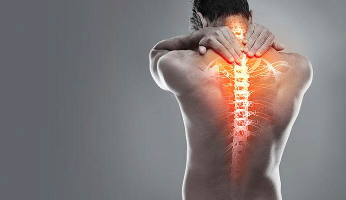
The symptoms associated with ulnar nerve compression may develop suddenly or gradually over a period of time. Symptoms may include an ache or pain at the back or inner aspect of the elbow and forearm, pins and needles, numbness or a burning sensation may also be present in a similar area or into the palm on the little finger side of the hand, the little finger and half of the ring finger. Tenderness and an increase in symptoms may be experienced when firmly pressing or tapping the nerve at the inner aspect of the elbow. Occasionally, in more severe cases, weakness of the hand and fingers may also be present.
In patients with inflammation contributing to the condition, symptoms may be worse at night or first thing in the morning. Occasionally, ulnar nerve compression may be associated with neck or upper back pain on the same side. In severe cases of ulnar nerve compression, obvious wasting of the muscles of the hand and forearm may be detected.
Diagnosis of ulnar nerve compression
A thorough subjective and objective examination from a physiotherapist may be sufficient to diagnose ulnar nerve compression. Nerve conduction studies may also be required to confirm diagnosis.
What is wrist tendonitis?
Wrist tendonitis is a relatively common overuse condition which may affect one or more wrist tendons and is characterized by inflammation and swelling of the affected tendons.
The forearm comprises of two long bones known as the radius and the ulna. These bones connect with several small bones known as the carpal bones forming the wrist (figure 1). Many muscles of the wrist and hand originate from the radius and ulnar and cross the wrist to insert into the bones of the wrist, hand and fingers via the wrist tendons. These muscles fall into two broad groups known as the wrist and finger flexors (which lie on the palm side of the forearm and wrist) and the wrist and finger extensors (which lie on the opposite side of the forearm and wrist) (figure 2). Generally, the wrist and finger flexors are responsible for bending the wrist and fingers (e.g. making a fist), whilst the wrist and finger extensors are responsible for straightening the wrist and fingers and bending them backwards. Collectively, the muscles are responsible for gripping activity, general movement and use of the wrist and fingers.
During contraction of the muscles of the wrist, tension is placed through the wrist tendons. When this tension is excessive due to too much repetition or high force, damage to the wrist tendons may occur. Wrist tendonitis is a condition whereby there is damage, with subsequent inflammation and degeneration to one or more of the wrist tendons. This is usually due to gradual wear and tear associated with overuse, however it may also occur traumatically due to a specific incident.
Causes of wrist tendonitis
Wrist tendonitis most commonly occurs due to repetitive or prolonged activities placing strain on the wrist tendons. These activities may include sports such as gymnastics, golf or racquet sports as well as manual work such as carpentry, painting, chopping wood, bricklaying, repetitive use of a hammer or screw driver, use of vibrating machinery, sewing and knitting or working at a computer. Wrist tendonitis may also occur from other activities involving forceful or repetitive gripping of the hand.
It is common for patients to develop this condition following a sudden increase in activities that place stress on the wrist tendons or due to a change in these activities. Occasionally, wrist tendonitis may develop suddenly. This can be due to a fall onto an outstretched hand or due to a forceful movement involving heavy lifting or a gripping force through the wrist. A history of wrist, elbow, shoulder, neck or upper back injury may increase the likelihood of a patient developing this condition.
Signs and symptoms of wrist tendonitis
The symptoms associated with this condition usually develop gradually over a period of time. Initially, symptoms may present as an ache or stiffness in the wrist and hand following an aggravating or unaccustomed activity. This may often be felt at night or first thing in the morning and may warm up with heat and movement in the early stages. As the condition progresses, pain may be felt with every day activities involving the wrist and fingers such as carrying groceries, opening jars, shaking hands or using the computer. Patients with wrist tendonitis may also experience swelling, crepitus or pain on firmly touching the affected wrist tendons.
Occasionally, pins and needles or numbness in the fingers may be experienced along with weakness in the fingers and hand. This may present as difficulty performing fine movements of the hand, reduced grip strength, or an increased frequency of dropping objects. Wrist tendonitis may also be associated with neck or upper back pain on the same side.
Diagnosis of wrist tendonitis
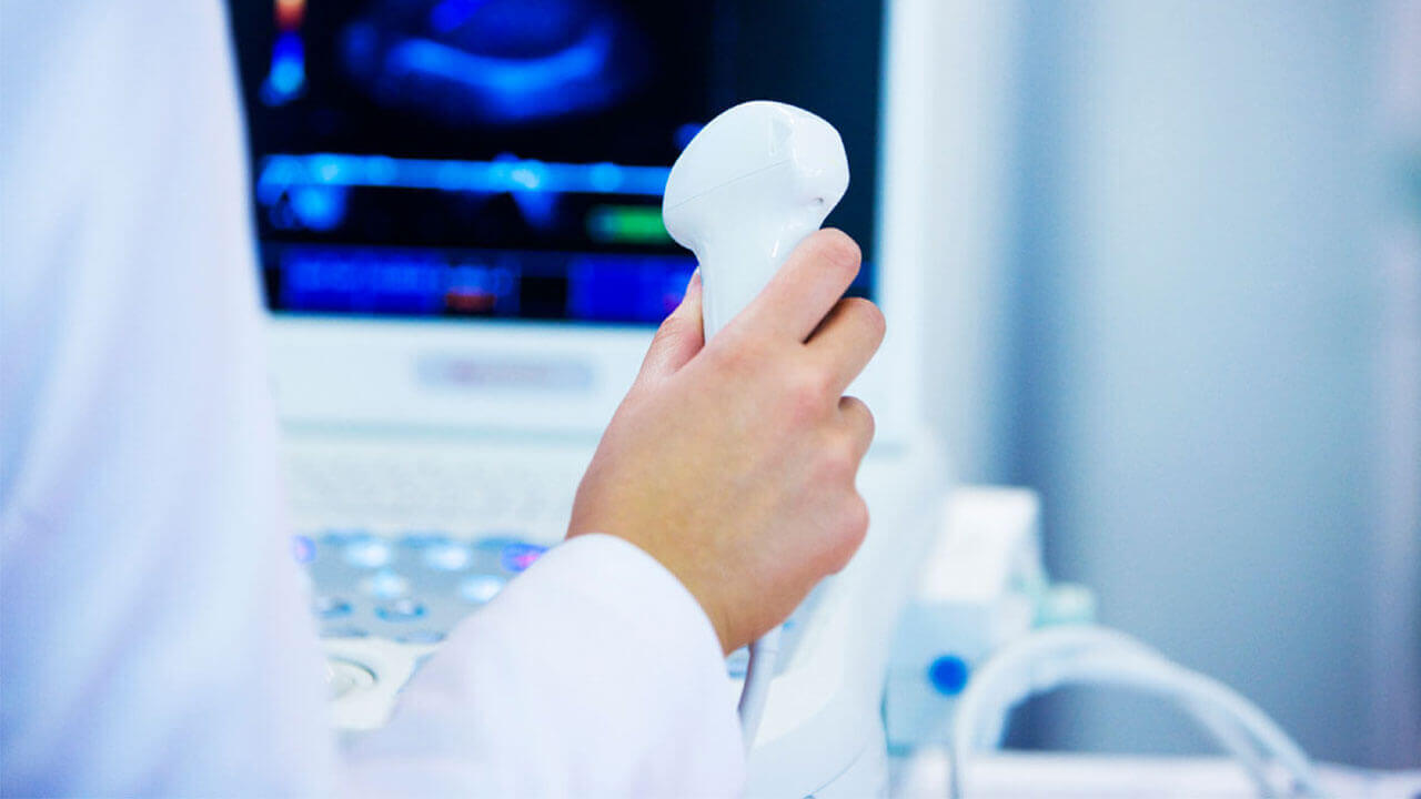
A thorough subjective and objective examination from a physiotherapist may be sufficient to diagnose wrist tendonitis. Further investigations such as an X-ray, Ultrasound, MRI or CT scan may be required to assist with diagnosis and assess the severity of the condition.
Diseases
-
Neck Pain
-
Shoulder & Elbow
-
Hand & Wrist
-
Arthritis
-
Lower Back Pain
-
Hip & Femur
-
Knee & Lower Leg
-
Foot & Ankle
-
Osteoporosis