Hip & Femur
The hip joint is one of the true ball-and-socket joints of the body. The hip socket is called the acetabulum and forms a deep cup that surrounds the ball of the upper thigh bone. The thigh bone itself is called the femur, and the ball on the end is the femoral head. The ball and socket arrangement gives the hip a large amount of motion needed for daily activities like walking, squatting, and stair-climbing.
The surfaces of the femoral head and the inside of the acetabulum are covered with articular cartilage. This material is about one-quarter of an inch thick in most large joints. Articular cartilage is a tough, slick material that allows the surfaces to slide against one another without damage.
The gluteus maximus is the largest of three gluteal muscles of the buttock. This muscle spans the side of the hip and joins the iliotibial band. The iliotibial band is a long tendon that passes over the bursa on the outside of the greater trochanter. It runs down the side of the thigh and attaches just below the outside edge of the knee. Two other buttock muscles attach to the greater trochanter, the gluteus medius and the gluteus minimus. These muscles are known as the abductors because they function to pull the lower leg away from the body – a motion that is called abduction. These muscles can be torn where they attach to the greater trochanter causing pain and and weakness as well as a snapping sensation.
Where friction must occur between muscles, tendons, and bones, there is usually a bursa. A bursa is a thin sac of tissue that contains a bit of fluid to lubricate the area where the friction occurs. The bursa is a normal structure, and the body will even produce a bursa in response to friction. The bursa next to the greater trochanter is called the greater trochanteric bursa.
The hip joint is surrounded by a water-tight pocket called the joint capsule. This capsule is formed by ligaments, connective tissue and synovial tissue. When the joint capsule is filled with sterile saline and is distended, the surgeon can insert the arthroscope into the pocket that is formed, turn on the lights and the camera and see inside the hip joint as if looking into an aquarium. The surgeon can see nearly everything that is inside the hip joint including: (1) the joint surfaces of the femoral head and acetabulum (2) the acetabular labrum and (3) the synovial lining of the joint.
A pelvic stress fracture is a condition characterized by an incomplete crack in one of the pelvic bones.
Each half of the pelvis comprises of the union of 3 bones known as the ischium, ilium and pubis (figure 1). Before puberty, these bones are separated by cartilage and eventually fuse to form one continuous bone by approximately 23 years of age. The two halves of the pelvis are called hemipelvises and together form the pelvis as a whole.
Many muscles of the hip, knee, lower back and abdomen attach to the pelvis. When these muscles contract, a pulling force is exerted on the bone. In addition, weight bearing activity places compressive forces on the pelvis via the hip joint. When these forces are excessive or too repetitive, and beyond what the bone can withstand, bony damage can gradually occur. This may initially result in a bony stress reaction, however, with continued damage may progress to a pelvic stress fracture.
The pubic ramus and ischium are two of the more commonly affected regions in a pelvic stress fracture. When these occur they are called ‘Pubic Ramus Stress Fracture’ and ‘Ischial Stress Fracture’ respectively.
Cause of a pelvic stress fracture
A pelvic stress fracture typically occurs over time with excessive weight bearing activity such as running, sprinting, jumping or dancing. They often occur following a recent increase in activity or change in training conditions (such as surface, footwear or technique changes etc) and are particularly common in long distance runners. Occasionally they may occur due to repetitive kicking in sports such as football or soccer. Pelvic stress fractures may also occur in women, following pregnancy, who commence excessive weight bearing activity without adequate pelvic and core stability.
Signs and symptoms of a pelvic stress fracture
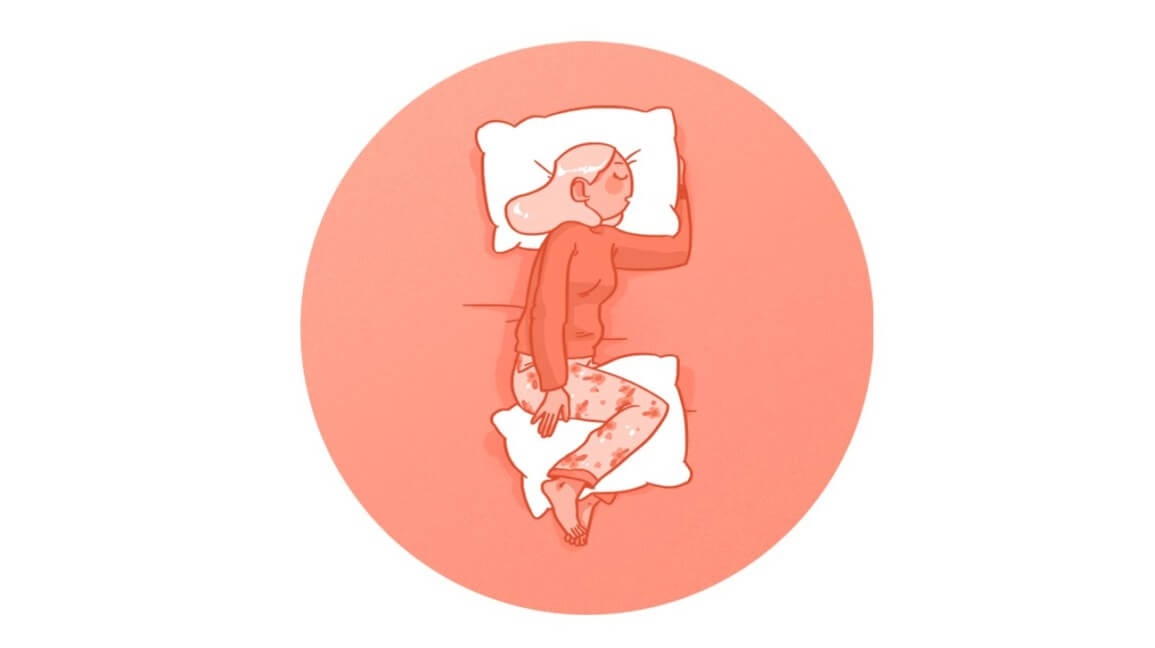
Patients with this condition typically experience a localized pain in the pelvic region that increases with impact activity (such as running, jumping, sprinting and hopping – often causing the patient to limp) and may decrease with rest. The pain may be severe enough that it causes the patient to stop the activity.
Depending on the location of the pelvic stress fracture, pain may be felt in the buttock, groin, hip or lower back and sometimes may radiate to the thigh or knee. In severe cases, walking or standing may be enough to aggravate symptoms. Other symptoms may include night ache, pain when resting or pain on firmly touching the affected region of the bone.
Diagnosis of a pelvic stress fracture
A thorough subjective and objective examination from a physiotherapist may be sufficient to diagnose a pelvic stress fracture. Further investigations such as an X-ray, MRI, CT scan or bone scan are usually required to confirm diagnosis and determine the severity of injury.
Prognosis of a pelvic stress fracture
With appropriate physiotherapy management, most patients with a pelvic stress fracture can make a full recovery (i.e. return to sport or normal activities) in a period of 3-12 months. In more severe cases, recovery may take 1-2 years, or longer, depending on the intervention required and a range of other factors. In rare cases, some patients may experience ongoing symptoms or complications which may require further management.
Osteitis pubis is an overuse injury characterized by tissue damage and inflammation to the pelvis at the site where the two pubic bones join (pubic symphysis), resulting in groin pain.
The pelvis is made up of two bones (hemipelvis). These two bones join together at the front of the pelvis at the pubic symphysis (figure 1). The pubic symphysis is made of cartilage which helps absorb forces between the two bones. There are several muscles that attach near the pubic symphysis (i.e. the adductors and abdominals). As these muscles contract (such as during running, kicking or performing sit-ups) a pulling force is exerted on the pubic symphysis. When these forces are excessive due to too much repetition or high force, damage to the pubic bones or symphysis may occur. When this results in inflammation to the pubic symphysis the condition is known as osteitis pubis.
Causes of osteitis pubis
Osteitis pubis is usually an overuse injury, which commonly occurs due to repetitive or prolonged activities placing strain on the pubic symphysis. This typically occurs due to repetitive running, kicking or change of direction activities. It is commonly seen in running sports such as football, hockey and athletics (particularly marathon runners).
Patients may also develop osteitis pubis due to excessive abdominal muscle contraction (such as during repetitive sit ups) or following inadequate rehabilitation of other injuries, such as adductor tendinopathy.
Signs and symptoms of osteitis pubis
Patients with osteitis pubis typically experience groin pain that develops gradually overtime. Pain may be experienced on one or both sides of the groin. Pain can sometimes also be experienced in the lower abdominals or at the front of the hips. Patients usually experience pain on firmly touching the pubic bone at the front of the pelvis (pubic symphysis – figure 1). Pain may also increase when squeezing the legs together or when moving the affected leg away from the midline of the body (abduction). Pain is usually aggravated by exercise such as running, kicking, performing sit-ups or change of direction activities.
In less severe cases of osteitis pubis, patients may only experience an ache or stiffness in the groin that increases upon rest following activity. These activities typically include running (particularly involving change of direction) and kicking. The pain associated with osteitis pubis may also warm up with activity in the initial stages of the condition.
As the condition progresses, patients may experience symptoms that increase during activity and affect performance. In severe cases the patient may be unable to continue the activity and may limp or ‘waddle’ as a result of the pain.
Diagnosis of osteitis pubis
A thorough subjective and objective examination from a physiotherapist is usually sufficient to diagnose osteitis pubis. Further investigations such as an X-ray, bone scan, MRI or CT scan may be required to assist with diagnosis and assess the severity of the condition
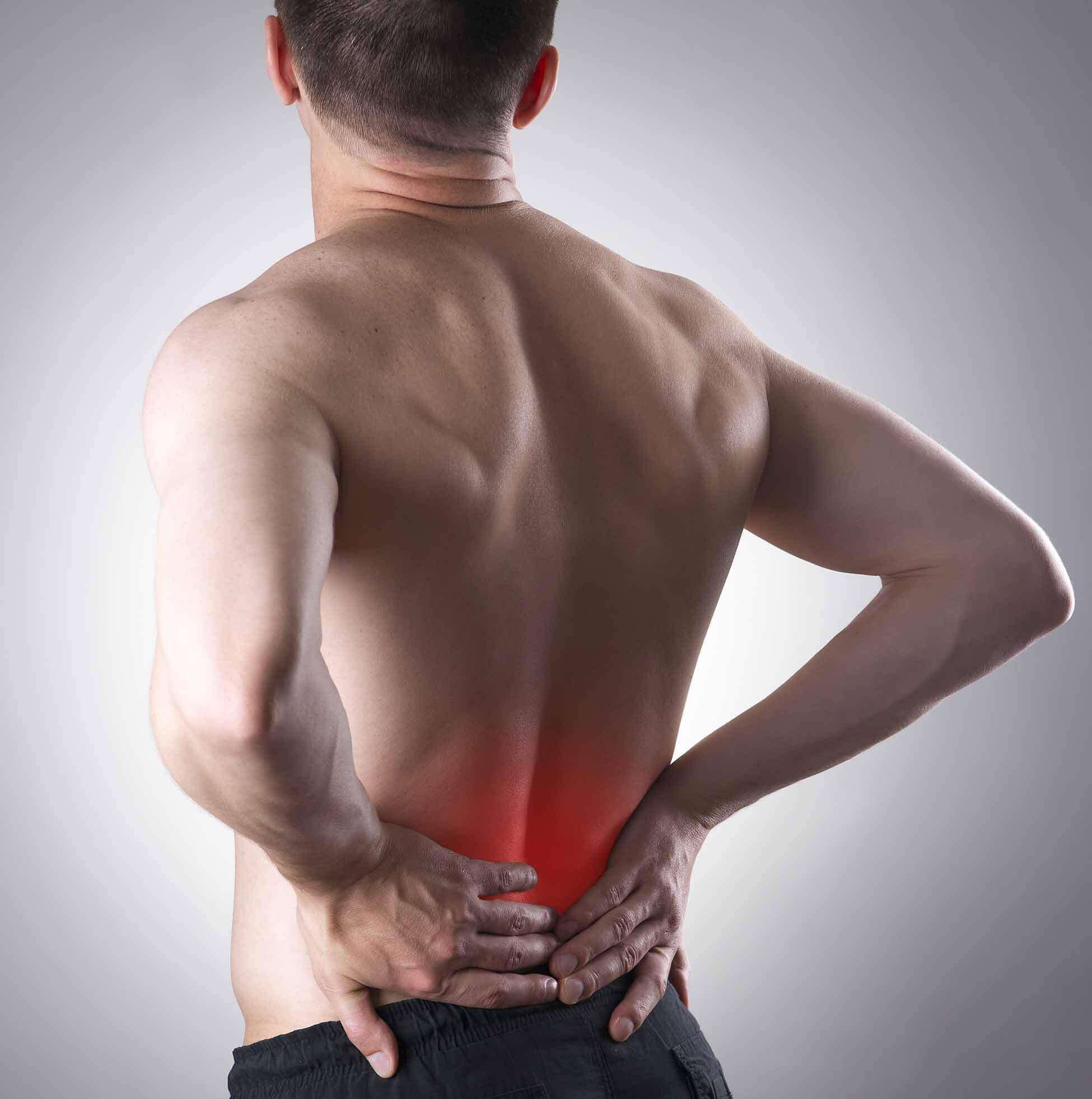
trochanteric bursitis of the Hip
Introduction
A common spot for bursitis is on the side of the hip. Here a large tendon passes over the bony bump on the side of the hip. The bony bump is called the greater trochanter. Inflammation in the bursa between the tendon and the greater trochanter is called trochanteric bursitis. This problem is common in older individuals. It may also occur in younger patients who are extremely active in exercises such as walking, running, or biking.
Anatomy
Where is the trochanteric bursa, and what does it do?
The hip joint is one of the true ball-and-socket joints of the body. The hip socket is called the acetabulum and forms a deep cup that surrounds the ball of the upper thigh bone (femur), or femoral head. Thick muscles of the buttock at the back and the thick muscles of the thigh in the front surround the hip.
The greater trochanter is the large bump on the outside of the upper end of the femur. This bump is the point where the large buttock muscles that move the hip connect to the femur. The gluteus maximus is the largest of these muscles. It attaches lower down on the femur.
Where friction occurs between muscles, tendons, and bones, there is usually a structure called a bursa. A bursa is a thin sac of tissue that contains fluid to lubricate the area and reduce friction. The bursa is a normal structure. The body will even produce a bursa in response to friction.
Causes
Why do I have this problem?
Sometimes a bursa can become inflamed (swollen and irritated) because of too much friction or because of an injury to the bursa. An inflamed bursa can cause pain because movement makes the structures around the bursa rub against it.
Friction can build in the bursa during walking if the long tendon on the side of the thigh is tight. It is unclear what causes this tightening of the tendon. The gluteus maximus attaches to this long tendon. As you walk, the gluteus maximus pulls this tendon over the greater trochanter with each step. When the tendon is tight, it rubs against the bursa. The rubbing causes friction to build in the bursa, leading to irritation and inflammation. Friction can also start if the outer hip muscle (gluteus medius) is weak, if one leg is longer than the other, or if you run on banked (slanted) surfaces.
Most cases of trochanteric bursitis appear gradually with no obvious underlying injury or cause. Trochanteric bursitis can occur after artificial replacement of the hip joint or other types of hip surgery. The cause may be a combination of changes in the way the hip works, the way it is aligned, or the way scar tissue has formed from the healing incision.
A fall on the hip can cause bleeding into the bursa, forming a hematoma. The bleeding is not serious, but the bursa may react to the blood by becoming inflamed. The inflammation causes the bursa to become thickened over time. This thickening, constant irritation, and inflammation may result in the condition becoming chronic, or long lasting.
Symptoms
What does the condition feel like?
The first symptom of trochanteric bursitis is usually pain. The pain can be felt in the area of the hip right over the bump that forms the greater trochanter. Eventually the pain may radiate down the outside of the thigh. As the problem progresses, the symptoms produce a limp when walking and stiffness in the hip joint. Eventually, the pain will also be present at rest and may even cause a problem with sleeping.
Diagnosis
How do doctors identify the problem?
The diagnosis of trochanteric bursitis begins with a history and physical examination. This is usually all that is necessary to make the diagnosis. Your doctor will want to know when the pain began and which motions cause the pain. A physical examination will be done to determine how much stiffness you have in the hip and if you have a limp. Once this is done, X-rays will most likely be ordered to make sure that there are no other abnormalities in the hip.
X-rays will usually not show trochanteric bursitis. If X-rays are suggested they are to rule out other problems that may be causing your hip pain. Sometimes it is difficult to tell whether the pain you are suffering is from trochanteric bursitis or underlying arthritis of the hip joint. An X-ray may give more information about the condition of the hip joint itself.
An injection of a local anesthetic into the bursa can help your doctor diagnose trochanteric bursitis. If the injection removes the pain immediately, then the diagnosis is probably trochanteric bursitis. Most physicians will also add a bit of cortisone medication to the novocaine to help treat the condition at the same time.
Introduction
Pain in the buttock that radiates down the leg is commonly called sciatica. The most common cause for sciatica is irritation of the spinal nerves in or near the lumbar spine. Sometimes the nerve irritation is not in the spine but further down the leg. One possible cause of sciatica is piriformis syndrome. Piriformis syndrome can be painful, but it is seldom dangerous and rarely leads to the need for surgery. Most people with this condition can reduce the pain and manage the problem with simple methods, such as physical therapy.
Anatomy
What parts of the body are involved?
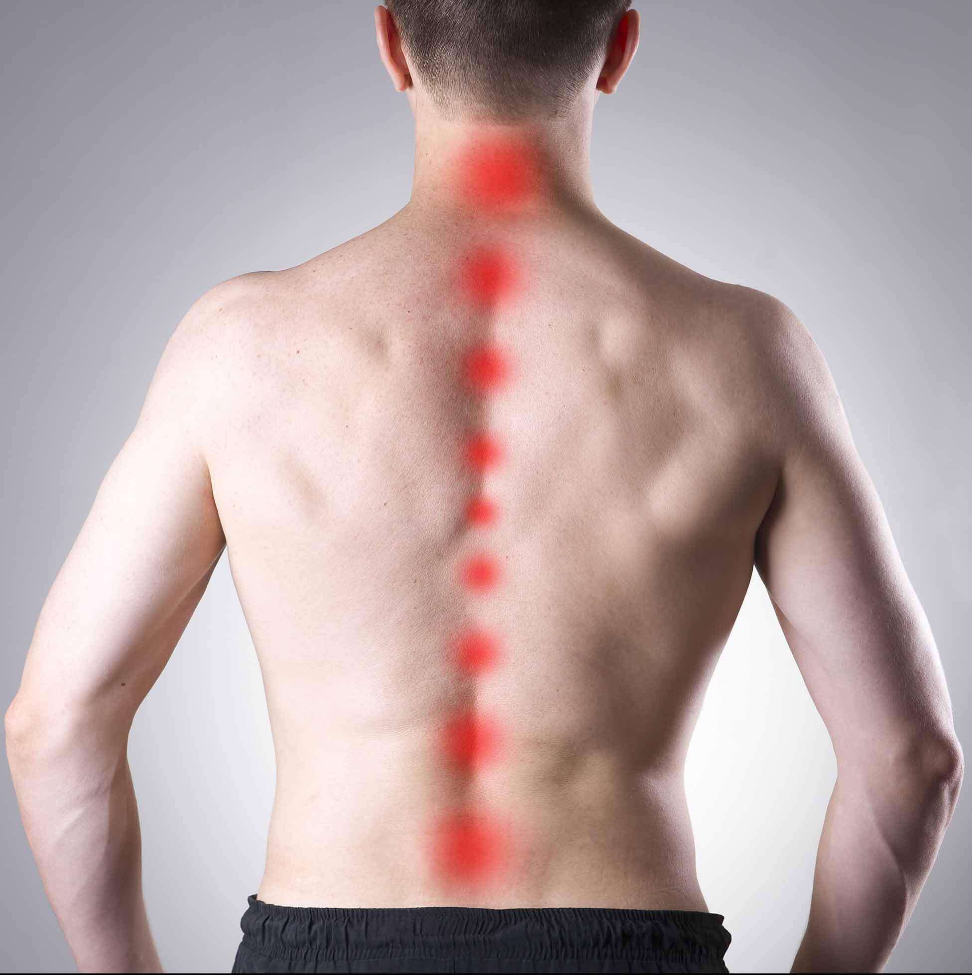
The lower lumbar spinal nerves leave the spine and join to form the sciatic nerve. The sciatic nerve leaves the pelvis through an opening called the sciatic notch.
The piriformis muscle begins inside the pelvis. It connects to the sacrum, the triangular shaped bone that sits between the pelvic bones at the base of the spine. The connection of the sacrum to the pelvis bones forms thesacroiliac joint. There is one sacroiliac joint on the left and one on the right of the low back. The other end of the piriformis muscle connects by a tendon to the greater trochanter, the bump of bone on the top side of your hip.
The piriformis muscle is one of the external rotators of the hip and leg. This means that as the muscle works, it helps to turn the foot and leg outward. Problems in the piriformis muscle can cause problems with the sciatic nerve. This is because the sciatic nerve runs under (and sometimes through) the piriformis muscle on its way out of the pelvis. The piriformis muscle can squeeze and irritate the sciatic nerve in this area, leading to the symptoms of sciatica.
Causes
What causes this problem?
The symptoms of sciatica come from irritation of the sciatic nerve. It’s still a mystery why the piriformis muscle sometimes starts to irritate the sciatic nerve. Many doctors think that the condition begins when the piriformis muscle goes into spasm and tightens against the sciatic nerve, squeezing the nerve against the bone of the pelvis.
In some cases, the muscle may be injured due to a fall onto the buttock. Bleeding in and around the piriformis muscle forms a hematoma. A hematoma describes the blood that has pooled in that area. The piriformis muscle begins to swell and put pressure on the sciatic nerve. Soon the hematoma dissolves, but the muscle goes into spasm.
The sciatic nerve stays irritated and continues to be a problem. Eventually the muscle heals, but some of the muscle fibers inside the piriformis muscle are replaced by scar tissue. Scar tissue is not nearly as flexible and elastic as normal muscle tissue. The piriformis muscle can tighten up and put constant pressure against the sciatic nerve.
Symptoms
What does the condition feel like?
Piriformis syndrome commonly causes pain that radiates down the back of the leg. The pain may be felt only on one side, though it is sometimes felt on both sides. The pain can radiate down the leg all the way to the foot and may be confused for a herniated disc in the lumbar spine. Changes in sensation and weakness in the leg or foot are rare. Some people say they feel a sensation of vague tingling down the leg.
Sitting may be difficult. Usually people with piriformis syndrome do not like to sit. When they do sit down, they tend to sit with the sore side buttock tilted up rather than sitting flat in the chair.
Diagnosis
How do doctors diagnose the problem?
Diagnosis begins with a complete history and physical exam. Your doctor will ask questions about your symptoms and how the pain is affecting your daily activities. Your doctor will also want to know what positions or activities make your symptoms worse or better. You will be asked about any injuries in the past and about any other medical problems you might have such as any arthritis that runs in the family.
Next the doctor examines you by checking your posture, how you walk, and where your pain is located. Your doctor checks to see which back movements cause pain or other symptoms.
Your skin sensation, muscle strength, and reflexes are also tested because it is difficult to distinguish pain coming from the sacroiliac joint from pain coming from other spine conditions.
If there is any question whether you might have an infection or some type of arthritis affecting multiple joints, laboratory tests may be ordered. You may need to have blood drawn and give a urine sample to send to the laboratory for special tests.
X-rays are commonly ordered of both the low back and pelvis. X-rays can give your doctor an idea about how much wear and tear has developed in the sacroiliac joint. X- rays of the lumbar spine and hips are also helpful to rule out problems in these areas that may look and act like sacroiliac joint problems.
Other radiological tests may also be useful. A magnetic resonance imaging (MRI) scan can be used to look at the lumbar spine and pelvis in much more detail and rule out other conditions in the area conditions. The MRI scan uses magnetic waves rather than X-rays and shows a very detailed picture of the soft tissues of the body.
A special type of MRI scan called neurography is being used more frequently to look at nerves. This uses a regular MRI scanner, but the computer settings are set to look for areas of irritation along a nerve. This may change the way doctors use the MRI to diagnose nerve problems such as piriformis syndrome, thoracic outlet syndrome, and carpal tunnel syndrome.
A bone scan is useful to see how the skeleton is reacting to any type of “stress,” such as an injury, an infection, or inflammation from arthritis. Chemical “tracers” are injected into your blood stream. The tracers then show up on special spine X- rays. The tracers collect in areas where the bone tissue is reacting strongly to some type of stress to the skeleton, such as arthritis and infection of the sacroiliac joint.
The most accurate way to tell if the piriformis muscle is the cause of pain is with a diagnostic injection into the muscle. The muscle is deep inside the buttock, so the injection requires X-ray guidance with a fluoroscope, a CT scanner, or an open MRI machine. Once the needle is placed in the muscle, an anesthetic can be injected into the muscle to paralyze the piriformis muscle. If the pain goes away after the injection, your doctor can be reasonably sure that the pain you are feel is from piriformis syndrome.
Ischiogluteal bursitis is a condition that causes pain in the buttock and is characterized by tissue damage and inflammation to the ischiogluteal bursa. A bursa is a small sac filled with lubricating fluid and is designed to reduce friction between adjacent soft tissue layers. The ischiogluteal bursa is located at the base of the pelvis, at the level of the bony prominence known as the ischial tuberosity (figure 1).
The hamstring muscles originate from the pelvis (ischial tuberosity) and insert into the top of the lower leg bones. The hamstring muscles attach to the pelvis via the hamstring tendon (figure 2). The ischiogluteal bursa lies between the hamstring tendon and the pelvic bone (ischial tuberosity).
The hamstring muscles are responsible for bending the knee and straightening the hip during activity and are particularly active during running, jumping and kicking. During contraction of the hamstrings, tension is placed through the hamstring tendon which in turn places friction on the ischiogluteal bursa. Pressure may also be placed on the ischiogluteal bursa during sitting. When these forces are excessive due to too much repetition or high force, irritation and inflammation of the ischiogluteal bursa may occur. This condition is known as an ischiogluteal bursitis.
Causes of ischiogluteal bursitis
Ischiogluteal bursitis most commonly occurs due to repetitive or prolonged activities placing strain on the ischiogluteal bursa. This typically occurs due to prolonged sitting (particularly on hard surfaces) or due to repetitive running, jumping or kicking activities (placing strain on the ischiogluteal bursa via the hamstring tendon). Occasionally, patients may develop this condition suddenly following a direct blow to the ischiogluteal bursa. This may occur due to a fall onto a hard surface.
Signs and symptoms of ischiogluteal bursitis
Patients with ischiogluteal bursitis typically experience pain in the lower buttock. In less severe cases, patients may only experience an ache or stiffness in the buttock that increases with rest following activities placing strain on the ischiogluteal bursa. These activities typically include sitting excessively (especially on hard surfaces), walking, running, jumping, kicking or climbing stairs. The pain associated with ischiogluteal bursitis may also warm up with activity in the initial stages of the condition.
As the condition progresses, patients may experience sharper or more severe symptoms that increase during sport or activity, affecting performance. Patients frequently experience pain on firmly touching the ischiogluteal bursa and hamstring tendon (figure 2). Occasionally, a feeling of lower limb weakness may also be present particularly when attempting to accelerate whilst running.
Diagnosis of ischiogluteal bursitis
A thorough subjective and objective examination from a physiotherapist may be sufficient to diagnose ischiogluteal bursitis. Further investigations such as an Ultrasound, X-ray, CT or MRI scan are often required to assist with diagnosis and assess the severity of the condition.
Osteonecrosis of the Hip
Sometimes called “wear-and-tear” arthritis, osteoarthritis is a common condition that many people develop during middle age or older. In 2011, more than 28 million people in the United States were estimated to have osteoarthritis. It can occur in any joint in the body, but most often develops in weight-bearing joints, such as the hip.
Osteoarthritis of the hip causes pain and stiffness. It can make it hard to do everyday activities like bending over to tie a shoe, rising from a chair, or taking a short walk.
Because osteoarthritis gradually worsens over time, the sooner you start treatment, the more likely it is that you can lessen its impact on your life. Although there is no cure for osteoarthritis, there are many treatment options to help you manage pain and stay active.
The hip is one of the body’s largest joints. It is a “ball-and-socket” joint. The socket is formed by the acetabulum, which is part of the large pelvis bone. The ball is the femoral head, which is the upper end of the femur (thighbone).
The bone surfaces of the ball and socket are covered with articular cartilage, a smooth, slippery substance that protects and cushions the bones and enables them to move easily.
The surface of the joint is covered by a thin lining called the synovium. In a healthy hip, the synovium produces a small amount of fluid that lubricates the cartilage and aids in movement
Description
Osteoarthritis is a degenerative type of arthritis that occurs most often in people 50 years of age and older, though it may occur in younger people, too.
In osteoarthritis, the cartilage in the hip joint gradually wears away over time. As the cartilage wears away, it becomes frayed and rough, and the protective joint space between the bones decreases. This can result in bone rubbing on bone. To make up for the lost cartilage, the damaged bones may start to grow outward and form bone spurs (osteophytes).
Osteoarthritis develops slowly and the pain it causes worsens over time.
A hip damaged by osteoarthritis.
Animation courtesy Visual Health Solutions, Inc.
Osteoarthritis has no single specific cause, but there are certain factors that may make you more likely to develop the disease, including:
- Increasing age
- Family history of osteoarthritis
- Previous injury to the hip joint
- Obesity
- Improper formation of the hip joint at birth, a condition known as developmental dysplasia of the hip
Even if you do not have any of the risk factors listed above, you can still develop osteoarthritis.
Symptoms
The most common symptom of hip osteoarthritis is pain around the hip joint. Usually, the pain develops slowly and worsens over time, although sudden onset is also possible. Pain and stiffness may be worse in the morning, or after sitting or resting for a while. Over time, painful symptoms may occur more frequently, including during rest or at night. Additional symptoms may include:
- Pain in your groin or thigh that radiates to your buttocks or your knee
- Pain that flares up with vigorous activity
- Stiffness in the hip joint that makes it difficult to walk or bend
- “Locking” or “sticking” of the joint, and a grinding noise (crepitus) during movement caused by loose fragments of cartilage and other tissue interfering with the smooth motion of the hip
- Decreased range of motion in the hip that affects the ability to walk and may cause a limp
- Increased joint pain with rainy weather
Diseases
-
Neck Pain
-
Shoulder & Elbow
-
Hand & Wrist
-
Arthritis
-
Lower Back Pain
-
Hip & Femur
-
Knee & Lower Leg
-
Foot & Ankle
-
Osteoporosis