Osteoporosis
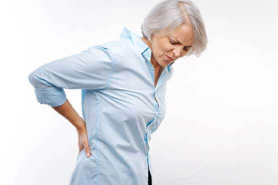
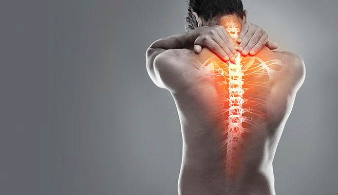
Osteoporosis is a disease of progressive bone loss associated with an increased risk of fractures. The term osteoporosis literally means porous bone. The disease often develops unnoticed over many years, with no symptoms or discomfort until a fracture occurs. Osteoporosis often causes a loss of height and dowager’s hump (a severely rounded upper back).
What Causes Osteoporosis?
Doctors have not identified a specific osteoporosis cause. They have identified risk factors that increase osteoporosis risk. According to the AAOS, these include:
advancing age
family history
unhealthy lifestyle choices
taking certain medications
Who Is at Risk For Osteoporosis?
Osteoporosis comes with a considerable number of risk factors. Some of them can be controlled while others cannot.
Risk factors you cannot control include:
having already experienced menopause
family history of osteoporosis
female gender, especially females of Caucasian or Asian origin
history of age-related height loss
history of broken bones
history of hormone-related medical conditions, such as hypothyroidism and Cushing’s disease
history of osteopenia
low body mass
age older than 50
Risk factors that are within your control include:
excess alcohol consumption
excess amounts of caffeine, protein and sodium in your diet
lack of calcium and vitamin D in your daily diet
lack of fruits and vegetables in your daily diet
lack of regular physical activity
long-term use of certain medications, including anticonvulsants and glucocorticoids
smoking/tobacco use
Talk to your doctor about your individual concerns and unique risk factors for osteoporosis. They can help you identify any medications that could be switched, if possible. Don’t stop treatments or medications without your doctor’s advice.
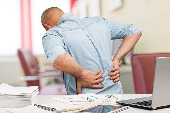
What Are the Symptoms of Osteoporosis?
Doctors call osteoporosis a “silent disease” because the condition often has no symptoms. The first sign a person has the condition is often a painful bone fracture.
Symptoms that could signal osteoporosis include progressive loss of height over time and a rounded upper back known as a dowager’s hump.
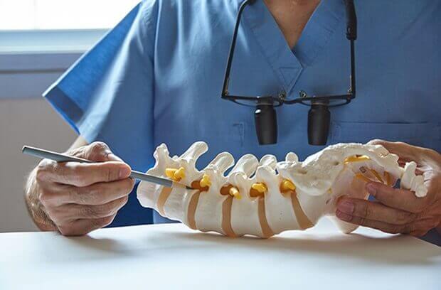
How Is Osteoporosis Diagnosed?
If you are at risk for osteoporosis or have experienced a bone fracture, a doctor may recommend a bone mineral density (BMD) test. A dual-energy X-ray absorptiometry (DEXA) scan is the most common BMD test. Similar to an X-ray, this painless test measures your bone density, especially at the hip and spine.
A DEXA scan can help determine how strong your bones are while letting your doctor monitor your bone density. This can confirm whether treatments are necessary and if so, whether or not they are working.
How Is Osteoporosis Treated?
Bone mass and skeletal structure generally cannot be replaced once it’s lost. Treatments for osteoporosis focus on retaining a person’s current bone mass level.
Several medical specialists may work together to treat the condition. These include an endocrinologist and an orthopedic specialist.
Treatments include the following..
Take steps to eat enough vitamin D and calcium. Both are bone-strengthening nutrients. According to the National Institutes of Health, male adults ages 19 to 70 and female adults ages 19 to 51 should consume at least 1,000 milligrams (mg) of calcium per day and 600 international units (IU) of vitamin D per day.
Women ages 51 and older should consume 1,200 mg of calcium per day. Calcium sources include low-fat cheese, milk and yogurt. Food manufacturers often add calcium and vitamin D to certain foods to enrich their nutritional values. Examples include orange juice, cereal, and bread.
Exercise stimulates bone tissue to grow stronger. This is only true for weight-bearing exercises. Examples include hiking, jogging, lifting weights and dancing. Exercises such as swimming and riding a bicycle may not be as effective. Falls cause a large percentage of osteoporosis-related injuries. Balance exercises such as tai chi can help prevent falls.
Refraining from smoking or drinking excessively can also prevent or at least delay osteoporosis. This means no more than one drink per day for women and one to two drinks per day for men
Osteoarthritis of the Elbow
Osteoarthritis of the elbow occurs when the cartilage surface of the elbow is damaged or becomes worn. This can happen because of a previous injury such as elbow dislocation or fracture. It may also be the result of degeneration of the joint cartilage from age. Osteoarthritis usually affects the weight-bearing joints, such as the hip and knee. The elbow is one of the least affected joints because of its well matched joint surfaces and strong stabilizing ligaments. As a result, the elbow joint can tolerate large forces across it without becoming unstable.
Diagnosis
X-ray showing an elbow with early degenerative changes.
A doctor can usually diagnose osteoarthritis of the elbow based on symptoms and standard X-rays. X-rays show the arthritic changes. Advanced diagnostic imaging, such as CT (computed tomography) or MRI (magnetic resonance imaging), is typically not needed to diagnose osteoarthritis of the elbow. Elbow osteoarthritis that occurs without previous injury is more common in men than women. Onset typically occurs in patients 50 years of age or older, but some patients can have symptoms earlier.
Most patients who are diagnosed with elbow osteoarthritis have a history of injury to the elbow, such as a fracture that involved the surface of the joint, or an elbow dislocation. The risk for elbow arthritis increases if:
- The patient needed surgery to repair the injury or reconstruct the joint
- There is loss of joint cartilage
- The joint surface cannot be repaired or reconstructed to its preinjury level
Injury to the ligaments resulting in an unstable elbow can also lead to osteoarthritis, even if the elbow surface is not damaged, because the normal forces across the elbow are altered, causing the joint to wear out more rapidly.
In some patients, no single injury to the elbow occurs. Work or outside activities can lead to osteoarthritis of the elbow if the patient places more demands on the joint than it can bear. For example, professional baseball pitchers place unusually high demands on their throwing elbows, which can lead to failure of the stabilizing ligaments. When this occurs, surgical reconstruction is usually needed. High-shear forces placed across the joint can lead to cartilage breakdown over a period of years.
The best way to prevent elbow arthritis is to avoid injury to the joint. When injury does occur, it is important to recognize it right away and get treatment. Individuals involved in heavy work or sports activities should maintain muscular strength around the elbow. Proper conditioning and technique should always be used.
Symptoms
The most common symptoms of elbow arthritis are:
- Pain
- Loss of range of motion
Both of these symptoms may not occur at the same time. Patients usually report a “grating” or “locking” sensation in the elbow. The “grating” is due to loss of the normal smooth joint surface. This is caused by cartilage damage or wear. The “locking” is caused by loose pieces of cartilage or bone that dislodge from the joint and become trapped between the moving joint surfaces, blocking motion.
Joint swelling may also occur, but this does not usually happen at first. Swelling occurs later, as the disease progresses.
In the later stages of osteoarthritis of the elbow, patients may notice numbness in their ring finger and small finger. This can be caused by elbow swelling or limited range of motion in the joint. The “funny bone” (ulnar nerve) is located in a tight tunnel behind the inner (medial) side of the elbow. Swelling in the elbow joint can put increased pressure on the nerve, causing tingling. If the elbow cannot be moved through its normal range of motion, it may stiffen into a position where it is bent (flexion). This can also cause pressure around the nerve to increase.
Osteonecrosis of the Hip
Sometimes called “wear-and-tear” arthritis, osteoarthritis is a common condition that many people develop during middle age or older. In 2011, more than 28 million people in the United States were estimated to have osteoarthritis. It can occur in any joint in the body, but most often develops in weight-bearing joints, such as the hip.
Osteoarthritis of the hip causes pain and stiffness. It can make it hard to do everyday activities like bending over to tie a shoe, rising from a chair, or taking a short walk.
Because osteoarthritis gradually worsens over time, the sooner you start treatment, the more likely it is that you can lessen its impact on your life. Although there is no cure for osteoarthritis, there are many treatment options to help you manage pain and stay active.
The hip is one of the body’s largest joints. It is a “ball-and-socket” joint. The socket is formed by the acetabulum, which is part of the large pelvis bone. The ball is the femoral head, which is the upper end of the femur (thighbone).
The bone surfaces of the ball and socket are covered with articular cartilage, a smooth, slippery substance that protects and cushions the bones and enables them to move easily.
The surface of the joint is covered by a thin lining called the synovium. In a healthy hip, the synovium produces a small amount of fluid that lubricates the cartilage and aids in movement.
The main problem in knee OA is degeneration of the articular cartilage. Articular cartilage is the smooth lining that covers the ends of bones where they meet to form the joint. The cartilage gives the knee joint freedom of movement by decreasing friction.
The articular cartilage is kept slippery by joint fluid made by the joint lining (the synovial membrane). The fluid, called synovial fluid, is contained in a soft tissue enclosure around synovial joints called the joint capsule.
An important substance present in articular cartilage and synovial fluid is called hyaluronic acid. Hyaluronic acid helps joints collect and hold water, improving lubrication and reducing friction. It also acts by allowing cells to move and work within the joint.
When the articular cartilage degenerates, or wears away, the bone underneath is uncovered and rubs against bone. Small outgrowths called bone spurs, or osteophytes, may form in the joint
Degeneration in a joint means the joint surfaces are starting to break down over time. The term degenerative arthritis is used by doctors to describe a condition where a joint wears out, usually over a period of many years. Some medical professionals call the condition osteoarthritis. Others use the term degenerative arthrosis. They prefer arthrosis because the term arthritis means inflammation. Degeneration by itself doesn’t always cause inflammation in the tissues of the joint. Still, these terms are generally used to mean the same thing.
What changes does osteoarthritis cause in the wrist joint?
The anatomy of the wrist joint is extremely complex, probably the most complex of all the joints in the body. The wrist is actually a collection of many joints and many bones. These joints and bones let us use our hands in many ways. The wrist must be extremely mobile to give our hands a full range of motion. At the same time, the wrist must provide the strength for heavy gripping.
The wrist is made up of eight separate small bones, called the carpal bones. The carpal bones connect the two bones of the forearm, the radius and the ulna, to the bones of the hand. The metacarpal bones are the long bones that lie underneath the palm. The metacarpals attach to the phalanges, which are the bones in the fingers and thumb.
One reason that the wrist is so complicated is because every small bone forms a joint with the bone next to it. This means that what we call the wrist joint is actually made up of many small joints. Ligaments connect all the small bones to each other. Ligaments also connect the bones of the wrist with the radius, ulna, and metacarpal bones.
Articular cartilage is the smooth, rubbery material that covers the bone surfaces in most joints. It protects the bone ends from friction when they rub together as the joint moves. Articular cartilage also acts sort of like a shock absorber. Damage to the articular cartilage eventually leads to osteoarthritis.
Related Document: A Patient’s Guide to Wrist Anatomy
Causes
How did I develop arthritis in my wrist?
Many wrist injuries, such as fractures and sprains, heal fairly easily. However, they can lead to problems much later in life. The injury changes the anatomy of the wrist just enough so that the parts no longer work smoothly together. The changes from the injury cause a lot of wear and tear on the wrist joint. Over time, this wear and tear degenerates the tissues of the joint, leading to wrist osteoarthritis. Doctors may also call this type of degeneration posttraumatic arthritis.
A bad sprain or fracture can actually damage the articular cartilage. The cartilage can also be bruised when too much pressure is put on the cartilage surface. The cartilage surface may not look any different. The injury often doesn’t show up until months later.
Sometimes the damage to the cartilage is severe. Pieces of the cartilage can actually be ripped away from the bone. These pieces do not grow back. Usually they must be surgically removed. If the pieces aren’t removed, they may float around in the joint, causing it to catch. They an also cause a lot of pain and do more damage to the joint surfaces.
Your body does not do a good job of repairing these holes in the cartilage surface. The holes fill up with scar tissue. Scar tissue is not as slick or rubbery as the articular cartilage.
Any kind of injury to the wrist joint can alter how the joint works. After a wrist fracture, the bone fragments may heal slightly differently. Ligament damage results in an unstable joint. Any time an injury changes the way the joint moves, even if the change is very subtle, the forces on the articular cartilage increase. It’s just like a machine; if the mechanism is out of balance, it wears out faster.
Over many years, this imbalance in joint mechanics can damage the articular cartilage. Since articular cartilage cannot heal itself very well, the damage adds up. Finally, the joint can no longer compensate for the damage, and your wrist begins to hurt.
Related Document: A Patient’s Guide to Ligament Injuries of the Wrist
Symptoms
What problems does arthritis of the wrist cause?
Pain is the main symptom of osteoarthritis of any joint. At first, the pain comes only with activity. Most of the time the pain lessens while doing the activity, but after stopping the activity the pain and stiffness increase. As the condition worsens, you may feel pain even when resting. The pain may interfere with sleep.
The wrist joints may be swollen. Your wrist may fill with fluid and feel tight, especially after use. When all the articular cartilage is worn off the joint surface, you may notice a squeaking sound when you move your wrist. Doctors call this creaking crepitus.
Osteoarthritis eventually affects the wrist’s motion. The wrist joint becomes stiff. Certain motions become painful. You may not be able to trust the joint when you lift objects in certain positions. This is because a pain reflex freezes the muscles when a joint is put in a position that causes pain. This happens without warning, and you can end up dropping whatever is in your hand.
Diagnosis
What tests will my doctor do?
The diagnosis of wrist osteoarthritis begins with a medical history. Your doctor will ask questions about your pain, how it interferes with your daily life, and whether anyone in your family has had similar problems. It is especially important to tell your doctor the details of any wrist injuries you’ve had, even if they happened many years ago.
Your doctor will then physically examine your wrist joint, and possibly other joints in your body. It may hurt when your doctor moves or probes your sore wrist. But it is important that your doctor sees how your wrist moves, how it is aligned, and exactly where it hurts.
You will probably need to have X-rays taken. X-rays are usually the best way to see what is happening with your bones. X-rays can help your doctor assess the damage and track how your joint changes over time. X-rays can also help your doctor estimate how much articular cartilage is left.
Your doctor may order blood tests if there is any question about the cause of your arthritis. Blood tests can show certain systemic diseases, such as rheumatoid arthritis
Introduction
Injuries of the ankle joint are common. While ankle fractures and ankle sprains heal pretty well, they can lead to problems much later in life. This is due to the wear and tear that occurs over the years after the injury. This condition is called osteoarthritis (OA) or posttraumatic arthritis. Trauma means injury, and the term posttraumatic arthritis is used to describe arthritis that develops after an injury.
How does the ankle joint work?
The ankle joint is made up of three bones: the lower end of the tibia (shinbone), the fibula (the small bone of the lower leg), and the talus (the bone that fits into the socket formed by the tibia and fibula).
The talus sits on top of the calcaneus (the heelbone). The talus moves mainly in one direction. It works like a hinge to allow your foot to move up and down.
Ligaments on both sides of the ankle joint help hold the bones together. Many tendons cross the ankle to move the ankle and the toes. (Ligaments connect bones to bones while tendons connect muscles to bones.) The large Achilles tendon in the back is the most powerful tendon in the foot. It connects the calf muscles to the heel bone and gives the foot the power for walking, running, and jumping.
Inside the joint, the bones are covered with a slick, smooth material called articular cartilage. Articular cartilage is the material that allows the bones to move against one another in the joints of the body. The cartilage lining is about one-quarter of an inch thick in most joints that carry body weight, such as the ankle, hip, or knee. It is soft enough to allow for shock absorption but tough enough to last a lifetime, as long as it is not injured.
Causes
Why do I have this problem?
OA is usually considered a type of degenerative arthritis, or wear-and-tear arthritis. Doctors consider OA pretty much the same whether it appears years after an injury to the joint or whether it appears without any history of injury. It behaves more or less the same way.
Over the past several years, there has been increasing evidence that OA is genetic, meaning that it runs in families. OA that occurs without any injury may prove to be related to differences in the chemical makeup of articular cartilage. People are born with these differences.
Injury to a joint, such as a bad sprain or fracture, can cause damage to the articular cartilage. The cartilage can be bruised when too much pressure is exerted on it. This damages the cartilage, although if you look at the surface it may not appear to be any different. The injury to the material doesn’t show up until months later. Sometimes the cartilage surface is damaged even more severely, and pieces of the cartilage are ripped from the bone. These pieces do not heal back and usually must be removed from the joint surgically. If not, they may float around in the joint, causing the joint to catch and be painful. These fragments of cartilage may also do more damage to the joint surface.
Once this cartilage is ripped away, it does not normally grow back. Unlike bone, holes in the surface are not simply replaced by the cartilage tissue around the hole. Instead the defects are filled with scar tissue. The scar tissue that forms is not nearly as good a material for covering joint surfaces as the cartilage it replaces. It just can’t support weight and isn’t smooth like true articular cartilage.
An injury to a joint, even if it does not injure the articular cartilage directly, can alter how the joint works. This is true for a fracture where the bone fragments heal differently from the way they were before the break occurred. It is also true when ligaments are damaged that lead to instability in the joint. When an injury results in a change in the way the joint moves, the injury may increase the forces on the articular cartilage. This is similar to any mechanical device or machinery. If the mechanism is out of balance, it wears out faster.
Over many years this imbalance in the joint mechanics can lead to damage to the articular surface. Since articular cartilage cannot heal itself very well, the damage adds up. Finally, the joint is no longer able to compensate for the increasing damage, and it begins to hurt. The damage occurs well before the pain begins.
In summary, arthritis may come from differences in how each of us is put together based on our genes, a condition best described as OA. Or arthritis may develop years after an injury that leads to slow damage to the joint surfaces, a condition probably best described as post-traumatic arthritis. Either way the joint is worn out, and it hurts. For the purposes of this document, we will refer to both types as OA.
Symptoms
What does arthritis of the ankle feel like?
Pain is the main problem with arthritis of any joint. This pain occurs at first only related to activity. Usually, once the activity gets underway there is not much pain, but after resting for several minutes the pain and stiffness increase. Later, when the condition worsens, pain may be present even at rest. The pain may interfere with sleep. The joint may swell, fill with fluid, and feel tight, especially following increased activity. As the articular cartilage starts to wear off the joint surface, the joint may squeak when moved. Doctors refer to this sound as crepitation.
OA will eventually affect the motion of a joint. The joint becomes stiff and loses flexibility. Certain movements can become painful, and it may become difficult to trust the joint to hold your weight in certain positions. The body has a pain reflex such that when a joint is put into a position that causes pain the muscles around the joint may stop working without warning. This reflex can cause a person to stumble or even fall when arthritis affects the ankle joint.
When OA has reached a very severe stage, the bone itself under the articular cartilage may become worn away. This can lead to increasing deformities around the joint. In the final stages, the alignment of the bones can begin to form odd angles where they meet at the joint.
Diagnosis
How do doctors identify OA?
The diagnosis of OA begins with a history of the problem. Details about any injuries that may have occurred to the joint, even years before, are important to understanding why the condition exists. Whether or not other family members have OA may shed some light on the problem.
Following the history, your doctor will examine the ankle joint and possibly other joints in your body. It will be important for your doctor to see how the motion of the ankle has been affected. The alignment of the ankle will be assessed. The nerves and circulation going to the legs and ankle will be checked. Your doctor will watch you walk to see if you have a noticeable limp.
Regular X-rays will be taken to see how severely the joint is damaged. This is usually the most important test to determine how bad the OA has become. How much articular cartilage is left in the ankle joint can be estimated with the X-rays.
If there is any question whether the arthritis may be coming from something other than OA, blood tests may be ordered to look for systemic diseases such as rheumatoid arthritis. A needle may be inserted into the joint to remove some of the joint fluid. This fluid may be sent to a lab to look for crystals due to gouty arthritis or signs of infection.
osteoarthritis acromioclavicular joint
Some joints in the body are more likely to develop problems from normal wear and tear. Degeneration causes the cartilage that cushions the joint to wear out. This type of arthritis is called osteoarthritis. Doctors sometimes refer to this type of arthritis as arthrosis.
The acromioclavicular (AC) joint in the shoulder is a common spot for osteoarthritis to develop in middle age. Degeneration of the AC joint can be painful and can cause difficulty using the shoulder for everyday activities.
Anatomy
What exactly is the AC joint?
The shoulder is made up of three bones: the scapula (shoulder blade), the humerus (upper arm bone), and the clavicle (collarbone).
The part of the scapula that makes up the roof of the shoulder and connects with the clavicle is called the acromion. The joint where the acromion and the clavicle join is the AC joint.
In some ways, the AC joint is like any other joint. It has two bones that need to connect but be flexible as well. The ends of the bones are covered with articular cartilage. Articular cartilage provides a slick, rubbery surface that allows the bones to glide over each other as you move. Cartilage also functions as sort of a shock absorber.
However, the AC joint is different from joints like the knee or ankle, because it doesn’t need to move very much. The AC joint only needs to be flexible enough for the shoulder to move freely. The AC joint just shifts a bit as the shoulder moves.
Causes
Why does degeneration of the AC joint occur?
We use our shoulder constantly. The resulting strain makes AC joint osteoarthritis a common disorder. The AC joint is under constant stress as the arm is used overhead. Weightlifters and others who repeatedly lift heavy amounts of weight overhead tend to have an increased incidence of the condition, and often at a younger age.
AC joint osteoarthritis may also develop following an injury to the joint, such as an AC joint separation. This injury is fairly common. A separation usually results from falling on the shoulder. The shoulder does heal, but many years later degeneration causes the AC joint to become painful.
Symptoms
What are the symptoms of this condition?
In its early stages, AC joint osteoarthritis usually causes pain and tenderness in the front of the shoulder around the joint. The pain is often worse when the arm is brought across the chest, since this motion compresses the joint. The pain is vague and may spread to include the shoulder, the front of the chest, and the neck. If the joint has been injured in the past, there may be a bigger bump over the joint on the affected shoulder than on the unaffected shoulder. The joint may also click or snap as it moves.
Diagnosis
What tests will my doctor do?
Your doctor will want to get a detailed medical history, including questions about your condition and how it is affecting you. You will need to answer questions about past injuries to your shoulder. You may be asked to rate your pain on a scale of one to ten. Your doctor will also want to know how much your pain affects your daily tasks.
Diagnosis of AC joint osteoarthritis is usually made by physical examination. The AC joint is usually tender. A key finding is pain as the joint is compressed. To test for this, your arm is pulled gently across your chest. Your doctor may inject a local anesthetic such as lidocaine into the joint. If the AC joint is the problem, the injection will temporarily reduce the pain.
Your doctor may want to take X-rays of the AC joint. X-rays can show narrowing of the joint and bone spurs around the joint, which are signs of degeneration.
Osteonecrosis of the Hip
Sometimes called “wear-and-tear” arthritis, osteoarthritis is a common condition that many people develop during middle age or older. In 2011, more than 28 million people in the United States were estimated to have osteoarthritis. It can occur in any joint in the body, but most often develops in weight-bearing joints, such as the hip.
Osteoarthritis of the hip causes pain and stiffness. It can make it hard to do everyday activities like bending over to tie a shoe, rising from a chair, or taking a short walk.
Because osteoarthritis gradually worsens over time, the sooner you start treatment, the more likely it is that you can lessen its impact on your life. Although there is no cure for osteoarthritis, there are many treatment options to help you manage pain and stay active.
The hip is one of the body’s largest joints. It is a “ball-and-socket” joint. The socket is formed by the acetabulum, which is part of the large pelvis bone. The ball is the femoral head, which is the upper end of the femur (thighbone).
The bone surfaces of the ball and socket are covered with articular cartilage, a smooth, slippery substance that protects and cushions the bones and enables them to move easily.
The surface of the joint is covered by a thin lining called the synovium. In a healthy hip, the synovium produces a small amount of fluid that lubricates the cartilage and aids in movement
Description
Osteoarthritis is a degenerative type of arthritis that occurs most often in people 50 years of age and older, though it may occur in younger people, too.
In osteoarthritis, the cartilage in the hip joint gradually wears away over time. As the cartilage wears away, it becomes frayed and rough, and the protective joint space between the bones decreases. This can result in bone rubbing on bone. To make up for the lost cartilage, the damaged bones may start to grow outward and form bone spurs (osteophytes).
Osteoarthritis develops slowly and the pain it causes worsens over time.
A hip damaged by osteoarthritis.
Animation courtesy Visual Health Solutions, Inc.
Osteoarthritis has no single specific cause, but there are certain factors that may make you more likely to develop the disease, including:
- Increasing age
- Family history of osteoarthritis
- Previous injury to the hip joint
- Obesity
- Improper formation of the hip joint at birth, a condition known as developmental dysplasia of the hip
Even if you do not have any of the risk factors listed above, you can still develop osteoarthritis.
Symptoms
The most common symptom of hip osteoarthritis is pain around the hip joint. Usually, the pain develops slowly and worsens over time, although sudden onset is also possible. Pain and stiffness may be worse in the morning, or after sitting or resting for a while. Over time, painful symptoms may occur more frequently, including during rest or at night. Additional symptoms may include:
- Pain in your groin or thigh that radiates to your buttocks or your knee
- Pain that flares up with vigorous activity
- Stiffness in the hip joint that makes it difficult to walk or bend
- “Locking” or “sticking” of the joint, and a grinding noise (crepitus) during movement caused by loose fragments of cartilage and other tissue interfering with the smooth motion of the hip
- Decreased range of motion in the hip that affects the ability to walk and may cause a limp
- Increased joint pain with rainy weather
Diseases
-
Neck Pain
-
Shoulder & Elbow
-
Hand & Wrist
-
Arthritis
-
Lower Back Pain
-
Hip & Femur
-
Knee & Lower Leg
-
Foot & Ankle
-
Osteoporosis