Shoulder & Elbow
Introduction
The shoulder is an elegant piece of machinery. It has the greatest range of motion of any joint in the body. However, this large range of motion can lead to joint problems.
Understanding how the different layers of the shoulder are built and connected can help you understand how the shoulder works, how it can be injured, and how challenging recovery can be when the shoulder is injured. The deepest layer of the shoulder includes the bones and the joints. The next layer is made up of the ligaments of the joint capsule. The tendons and the muscles come next.
Important Structures
The important structures of the shoulder can be divided into several categories. These include
- bones and joints
- ligaments and tendons
- muscles
- nerves
- blood vessels
- bursae
Bones and Joints
The bones of the shoulder are the humerus (the upper arm bone), the scapula (the shoulder blade), and the clavicle (the collar bone). The roof of the shoulder is formed by a part of the scapula called the acromion.
There are actually four joints that make up the shoulder. The main shoulder joint, called the glenohumeral joint, is formed where the ball of the humerus fits into a shallow socket on the scapula. This shallow socket is called the glenoid.
The acromioclavicular (AC) joint is where the clavicle meets the acromion. The sternoclavicular (SC) joint supports the connection of the arms and shoulders to the main skeleton on the front of the chest.
A false joint is formed where the shoulder blade glides against the thorax (the rib cage). This joint, called the scapulothoracic joint, is important because it requires that the muscles surrounding the shoulder blade work together to keep the socket lined up during shoulder movements.
Articular cartilage is the material that covers the ends of the bones of any joint. Articular cartilage is about one-quarter of an inch thick in most large, weight-bearing joints. It is a bit thinner in joints such as the shoulder, which don’t normally support weight. Articular cartilage is white and shiny and has a rubbery consistency. It is slippery, which allows the joint surfaces to slide against one another without causing any damage. The function of articular cartilage is to absorb shock and provide an extremely smooth surface to make motion easier. We have articular cartilage essentially everywhere that two bony surfaces move against one another, or articulate. In the shoulder, articular cartilage covers the end of the humerus and socket area of the glenoid on the scapula
Ligaments and Tendons
There are several important ligaments in the shoulder. Ligaments are soft tissue structures that connect bones to bones. A joint capsule is a watertight sac that surrounds a joint. In the shoulder, the joint capsule is formed by a group of ligaments that connect the humerus to the glenoid. These ligaments are the main source of stability for the shoulder. They help hold the shoulder in place and keep it from dislocating.
Ligaments attach the clavicle to the acromion in the AC joint. Two ligaments connect the clavicle to the scapula by attaching to the coracoid process, a bony knob that sticks out of the scapula in the front of the shoulder.
A special type of ligament forms a unique structure inside the shoulder called the labrum. The labrum is attached almost completely around the edge of the glenoid. When viewed in cross section, the labrum is wedge-shaped. The shape and the way the labrum is attached create a deeper cup for the glenoid socket. This is important because the glenoid socket is so flat and shallow that the ball of the humerus does not fit tightly. The labrum creates a deeper cup for the ball of the humerus to fit into.
The labrum is also where the biceps tendon attaches to the glenoid. Tendons are much like ligaments, except that tendons attach muscles to bones. Muscles move the bones by pulling on the tendons. The biceps tendon runs from the biceps muscle, across the front of the shoulder, to the glenoid. At the very top of the glenoid, the biceps tendon attaches to the bone and actually becomes part of the labrum. This connection can be a source of problems when the biceps tendon is damaged and pulls away from its attachment to the glenoid.
The tendons of the rotator cuff are the next layer in the shoulder joint. Four rotator cuff tendons connect the deepest layer of muscles to the humerus.
Muscles
The rotator cuff tendons attach to the deep rotator cuff muscles. This group of muscles lies just outside the shoulder joint. These muscles help raise the arm from the side and rotate the shoulder in the many directions. They are involved in many day-to-day activities. The rotator cuff muscles and tendons also help keep the shoulder joint stable by holding the humeral head in the glenoid socket.
The large deltoid muscle is the outer layer of shoulder muscle. The deltoid is the largest, strongest muscle of the shoulder. The deltoid muscle takes over lifting the arm once the arm is away from the side.
Nerves
The main nerves that travel into the arm run through the axilla under the shoulder. Three main nerves begin together at the shoulder: the radial nerve, the ulnar nerve, and the median nerve. These nerves carry the signals from the brain to the muscles that move the arm. The nerves also carry signals back to the brain about sensations such as touch, pain, and temperature. There is also an important nerve that travels around the back of the shoulder joint to supply sensation to a small area of skin on the outside of the shoulder and motor signals to the deltoid muscle. This nerve is called the axillary nerve.
Blood Vessels
Traveling along with the nerves are the large vessels that supply the arm with blood. The large axillary artery travels through the axilla. If you place your hand in your armpit, you may be able to feel the pulsing of this large artery. The axillary artery has many smaller branches that supply blood to different parts of the shoulder. The shoulder has a very rich blood supply.
Bursae
Sandwiched between the rotator cuff muscles and the outer layer of large bulky shoulder muscles are structures known as bursae. Bursae are everywhere in the body. They are found wherever two body parts move against one another and there is no joint to reduce the friction. A single bursa is simply a sac between two moving surfaces that contains a small amount of lubricating fluid.
Think of a bursa like this: If you press your hands together and slide them against one another, you produce some friction. In fact, when your hands are cold you may rub them together briskly to create heat from the friction. Now imagine that you hold in your hands a small plastic sack that contains a few drops of salad oil. This sack would let your hands glide freely against each other without a lot of friction.
Summary
As you can see, the shoulder is extremely complex, with a design that provides maximum mobility and range of motion. Besides big lifting jobs, the shoulder joint is also responsible for getting the hand in the right position for any function. When you realize all the different ways and positions we use our hands every day, it is easy to understand how hard daily life can be when the shoulder isn’t working well.
Introduction
At first, the elbow seems like a simple hinge. But when the complexity of the interaction of the elbow with the forearm and wrist is understood, it is easy to see why the elbow can cause problems when it does not function correctly. Part of what makes us human is the way we are able to use our hands. Effective use of our hands requires stable, painless elbow joints.
Important Structures
The important structures of the elbow can be divided into several categories. These include
- bones and joints
- ligaments and tendons
- muscles
- nerves
- blood vessels
Bones and Joints
The bones of the elbow are the humerus (the upper arm bone), the ulna (the larger bone of the forearm, on the opposite side of the thumb), and the radius (the smaller bone of the forearm on the same side as the thumb). The elbow itself is essentially a hinge joint, meaning it bends and straightens like a hinge. But there is a second joint where the end of the radius (the radial head) meets the humerus. This joint is complicated because the radius has to rotate so that you can turn your hand palm up and palm down. At the same time, it has to slide against the end of the humerus as the elbow bends and straightens. The joint is even more complex because the radius has to slide against the ulna as it rotates the wrist as well. As a result, the end of the radius at the elbow is shaped like a smooth knob with a cup at the end to fit on the end of the humerus. The edges are also smooth where it glides against the ulna.
Articular cartilage is the material that covers the ends of the bones of any joint. Articular cartilage can be up to one-quarter of an inch thick in the large, weight-bearing joints. It is a bit thinner in joints such as the elbow, which don’t support weight. Articular cartilage is white, shiny, and has a rubbery consistency. It is slippery, which allows the joint surfaces to slide against one another without causing any damage.
The function of articular cartilage is to absorb shock and provide an extremely smooth surface to make motion easier. We have articular cartilage essentially everywhere that two bony surfaces move against one another, or articulate. In the elbow, articular cartilage covers the end of the humerus, the end of the radius, and the end of the ulna.
Ligaments and Tendons
There are several important ligaments in the elbow. Ligaments are soft tissue structures that connect bones to bones. The ligaments around a joint usually combine together to form a joint capsule. A joint capsule is a watertight sac that surrounds a joint and contains lubricating fluid called synovial fluid.
In the elbow, two of the most important ligaments are the medial collateral ligament and the lateral collateral ligament. The medial collateral is on the inside edge of the elbow, and the lateral collateral is on the outside edge. Together these two ligaments connect the humerus to the ulna and keep it tightly in place as it slides through the groove at the end of the humerus. These ligaments are the main source of stability for the elbow. They can be torn when there is an injury or dislocation to the elbow. If they do not heal correctly the elbow can be too loose, or unstable.
There is also an important ligament called the annular ligament that wraps around the radial head and holds it tight against the ulna. The word annular means ring shaped, and the annular ligament forms a ring around the radial head as it holds it in place. This ligament can be torn when the entire elbow or just the radial head is dislocated.
There are several important tendons around the elbow. The biceps tendon attaches the large biceps muscle on the front of the arm to the radius. It allows the elbow to bend with force. You can feel this tendon crossing the front crease of the elbow when you tighten the biceps muscle
The triceps tendon connects the large triceps muscle on the back of the arm with the ulna. It allows the elbow to straighten with force, such as when you perform a push-up.
The muscles of the forearm cross the elbow and attach to the humerus. The outside, or lateral, bump just above the elbow is called the lateral epicondyle. Most of the muscles that straighten the fingers and wrist all come together in one tendon to attach in this area. The inside, or medial, bump just above the elbow is called the medial epicondyle. Most of the muscles that bend the fingers and wrist all come together in one tendon to attach in this area. These two tendons are important to understand because they are a common location of tendonitis.
Muscles
The main muscles that are important at the elbow have been mentioned above in the discussion about tendons. They are the biceps, the triceps, the wrist extensors (attaching to the lateral epicondyle) and the wrist flexors (attaching to the medial epicondyle).
Nerves
All of the nerves that travel down the arm pass across the elbow. Three main nerves begin together at the shoulder: the radial nerve, the ulnar nerve, and the median nerve. These nerves carry signals from the brain to the muscles that move the arm. The nerves also carry signals back to the brain about sensations such as touch, pain, and temperature.
Some of the more common problems around the elbow are problems of the nerves. Each nerve travels through its own tunnel as it crosses the elbow. Because the elbow must bend a great deal, the nerves must bend as well. Constant bending and straightening can lead to irritation or pressure on the nerves within their tunnels and cause problems such as pain, numbness, and weakness in the arm and hand.
Blood Vessels
Traveling along with the nerves are the large vessels that supply the arm with blood. The largest artery is the brachial artery that travels across the front crease of the elbow. If you place your hand in the bend of your elbow, you may be able to feel the pulsing of this large artery. The brachial artery splits into two branches just below the elbow: the ulnar artery and the radial artery that continue into the hand. Damage to the brachial artery can be very serious because it is the only blood supply to the hand.
Introduction
Shoulder instability means that the shoulder joint is too loose and is able to slide around too much in the socket. In some cases, the unstable shoulder actually slips out of the socket. If the shoulder slips completely out of the socket, it has become dislocated. If not treated, instability can lead to arthritis of the shoulder joint.
Anatomy
What parts of the shoulder are involved?
The shoulder is made up of three bones: the scapula (shoulder blade), the humerus (upper arm bone,) and the clavicle (collarbone).
The rotator cuffconnects the humerus to the scapula. The rotator cuff is actually made up of the tendons of four muscles: the supraspinatus, infraspinatus, teres minor, and subscapularis.
Tendons attach muscles to bones. Muscles move bones by pulling on tendons. The muscles of the rotator cuff also keep the humerus tightly in the socket. A part of the scapula, called the glenoid, makes up the socket of the shoulder. The glenoid is very shallow and flat. A rim of soft tissue, called the labrum, surrounds the edge of the glenoid, making the socket more like a cup. The labrum turns the flat surface of the glenoid into a deeper socket that molds to fit the head of the humerus.
Surrounding the shoulder joint is a watertight sac called the joint capsule. The joint capsule holds fluids that lubricate the joint. The walls of the joint capsule are made up of ligaments. Ligaments are soft connective tissues that attach bones to bones. The joint capsule has a considerable amount of slack, loose tissue, so that the shoulder is unrestricted as it moves through its large range of motion. If the shoulder moves too far, the ligaments become tight and stop any further motion, sort of like a dog coming to the end of its leash.
Dislocations happen when a force overcomes the strength of the rotator cuff muscles and the ligaments of the shoulder. Nearly all dislocations are anterior dislocations, meaning that the humerus slips out of the front of the glenoid. Only three percent of dislocations are posterior dislocations, or out the back.
Sometimes the shoulder does not come completely out of the socket. It slips only partially out and then returns to its normal position. This is called subluxation.
Causes
What makes a shoulder become unstable?
Shoulder instability often follows an injury that caused the shoulder to dislocate. This initial injury is usually fairly significant, and the shoulder must be reduced. To reduce a shoulder means it must be manually put back into the socket. The shoulder may seem to return to normal, but the joint often remains unstable. The ligaments that hold the shoulder in the socket, along with the labrum (the cartilage rim around the glenoid), may have become stretched or torn. This makes them too loose to keep the shoulder in the socket when it moves in certain positions. An unstable shoulder can result in repeated episodes of dislocation, even during normal activities. Instability can also follow less severe shoulder injuries.
In some cases, shoulder instability can happen without a previous dislocation. People who do repeated shoulder motions may gradually stretch out the joint capsule. This is especially common in athletes such as baseball pitchers, volleyball players, and swimmers. If the joint capsule gets stretched out and the shoulder muscles become weak, the ball of the humerus begins to slip around too much within the shoulder. Eventually this can cause irritation and pain in the shoulder.
A genetic problem with the connective tissues of the body can lead to ligaments that are too elastic. When ligaments stretch too easily, they may not be able to hold the joints in place. All the joints of the body may be too loose. Some joints, such as the shoulder, may be easily dislocated. People with this condition are sometimes referred to as double-jointed.
Symptoms
What problems does an unstable shoulder cause?
Chronic instability causes several symptoms. Frequent subluxation is one. In subluxation, the shoulder may slip (sublux) in certain positions, and the shoulder may actually feel loose. This commonly happens when the hand is raised above the head, for example while throwing. Subluxation of the shoulder usually causes a quick feeling of pain, like something is slipping or pinching in the shoulder. Over time, you may stop using the shoulder in ways that cause subluxation.
The shoulder may become so loose that it starts to dislocate frequently. This can be a real problem, especially if you can’t get it back in the socket and must go to the emergency room every time. A shoulder dislocation is usually very obvious. The injury is very painful, and the shoulder looks abnormal. Any attempted shoulder movements cause extreme pain. A dislocated shoulder can damage the nerves around the shoulder joint.
If the nerves have been stretched, a numb spot may develop on the outside of the arm, just below the top point of the shoulder. Several of the shoulder muscles may become slightly weak until the nerve recovers. But the weakness is usually temporary.
Diagnosis
What tests will my doctor run?
Your doctor will diagnose shoulder instability primarily through your medical history and physical exam. The medical history will include many questions about past shoulder injuries, your pain, and the ways your symptoms are affecting your activities.
In the physical exam, your doctor will feel and move your shoulder, checking it for strength and mobility. Your doctor will stress the shoulder to test the ligaments. When the shoulder is stretched in certain directions, you may get the feeling that the shoulder is going to dislocate. This is a very important sign of instability. It is called an apprehension sign. (Don’t worry. Unless your shoulder is extremely loose, it will not dislocate.)
Your doctor may order an X-ray. X-rays can help confirm that your shoulder was dislocated or injured in the past.
If your doctor is unsure about the diagnosis, you may need to undergo further tests. A surgeon may need to examine your shoulder using an arthroscope while you are under general anesthesia, which puts you to sleep. An arthroscope is a tiny TV camera inserted into the shoulder through a small incision. This allows a good look at the muscles and ligaments of the shoulder. When you are awake, it is hard to test the ligaments because you automatically tighten the muscles during the exam.
When you go to the doctor with a dislocated shoulder, X-rays are necessary to rule out a fracture. X-rays are usually done after the shoulder is put back into joint. This allows your doctor to make sure the joint is back in place
A dislocated shoulder is a common traumatic sporting injury characterized by tearing of the connective tissue surrounding the shoulder joint with subsequent displacement of the bones forming the joint so they are no longer situated next to each other (i.e. the upper arm bone comes out of the socket).
The shoulder joint is a ball and socket joint. The shoulder blade gives rise to the socket of the shoulder, whilst the ball of the shoulder arises from the top of the humerus (upper arm bone). Surrounding the ball and socket joint is strong connective tissue holding the bones together known as the shoulder joint capsule (figure 1). In addition, the shoulder joint ligaments (glenohumeral ligaments) help reinforce the joint capsule and a group of muscles known as the rotator cuff cross the shoulder joint and actively help to hold the shoulder joint in position increasing the shoulder’s stability.
During certain movements of the arm, stretching forces are applied to the shoulder joint capsule. When these forces are excessive and beyond what the shoulder can withstand, tearing of the connective tissue may occur. This may allow the upper arm bone (humerus) to move out of the socket if the forces involved are too great and beyond what the connective tissue and supporting muscles can withstand. When this occurs, the condition is known as a dislocated shoulder.
Dislocated shoulders are commonly seen in contact sports such as rugby or Australian rules football, high velocity sports, such as downhill skiing, snowboarding, skateboarding, or in competitive overhead sports such as basketball or water polo.
Causes of a dislocated shoulder
A dislocated shoulder typically occurs traumatically due to forces pushing the ball shaped aspect of the upper arm bone out of the socket. This may occur due to a direct impact or more commonly, due to a fall on the outstretched arm. The usual movements involved in this injury are a combination of shoulder abduction (side elevation) and excessive external rotation (outer rotation of the humerus) (figure 2). This is often combined with a force to the back of the shoulder or front of the elbow (or both). The result is most commonly an anterior shoulder dislocation (the ball moving forwards out of the socket). Other shoulder movements, positions or external forces, however, can result in an inferior or posterior dislocation of the shoulder (i.e. the ball moving below or backwards out of the socket), however, these tend to be less common.
Occasionally, a dislocated shoulder may occur with a relatively trivial movement (such as sneezing or turning over in bed) in these cases, there is usually a history of previous traumatic shoulder dislocation, shoulder instability or generalized ligamentous laxity throughout the body.
Signs and symptoms of a dislocated shoulder
Patients with a dislocated shoulder usually experience sudden severe pain at the time of injury. The pain is usually so intense that the patient cannot continue activity and will often cradle the arm against their body. Pain is usually felt in the shoulder region, however, can occasionally radiate down the arm.
Patients will often experience a sensation of the shoulder ‘popping out’ at the time of injury. Visible deformity of the shoulder joint may also be detected when compared to the other side. Occasionally, patients may experience pins and needles or numbness in the shoulder, arm or hand.
Once a dislocated shoulder has been ‘relocated’ (i.e. the ball positioned back in the socket by a sports medicine professional), patients may experience a feeling of weakness in the shoulder and an ache that may increase to a sharper pain with certain movements. These symptoms generally resolve over time with appropriate rehabilitation. However, patients with this condition may be left with a feeling of impending dislocation when the shoulder is placed in certain positions, particularly the combination of abduction and external rotation (figure 2).
Diagnosis of a dislocated shoulder
A thorough subjective and objective examination from a physiotherapist is usually sufficient to diagnose a dislocated shoulder. X-ray investigation is usually required to determine if there are any fractures associated with the dislocation and to confirm diagnosis. Further investigations such as a CT scan, Ultrasound or MRI may be required to assist diagnosis and determine involvement of other structures.
Introduction
The scapulothoracic joint is located where the shoulder blade (also called the scapula) glides along the chest wall (the thorax). When movement of this joint causes feelings or sounds of grating, grinding, popping, or thumping, doctors call it snapping scapula syndrome.
Snapping scapula syndrome is fairly rare. When it happens, the soft tissues between the scapula and the chest wall are thick, irritated, or inflamed. Snapping scapula syndrome can also happen if the bones of the shoulder blade or rib cage grate over one another.
Anatomy
What parts of the body are involved in this condition?
The shoulder is made up of three bones: the humerus (upper arm bone), the clavicle (collarbone), and the scapula (shoulder blade). Two large muscles attach to the front part of the scapula where it rests against the chest wall. One of them, called the subscapularis muscle, attaches over the front of the scapula where it faces the chest wall. The serratus anterior muscle attaches along the edge of the scapula nearest the spine. It passes in front of the scapula, wraps around the chest wall, and connects to the ribs on the front part of the chest.
A bursa is a fluid-filled sac that cushions body tissues from friction. A bursa sits between the two muscles of the scapula. There is also a bursa in the space between the serratus anterior muscle and the chest wall. When bursa sacs become inflamed, the condition is called bursitis.
Scapulothoracic bursitis refers to inflammation in the bursa under the shoulder blade. This type of bursitis is most common in the upper corner of the scapula nearest the spine. It also occurs under the lower tip of the scapula. In either case, it can cause the sounds and sensations of snapping scapula syndrome. A person can have bursitis in the joint without any grinding or popping.
Causes
What causes this condition?
Snapping scapula is caused by problems in the soft tissues or bones of the scapula and chest wall. It can start when the tissues between the scapula and shoulder blade thicken from inflammation. The inflammation is usually caused by repetitive movements. Certain motions of the shoulder done over and over again, such as the movements of pitching baseballs or hanging wallpaper, can cause the tissues of the joint to become inflamed.
In other cases, the muscles under the scapula have shrunk (atrophied) from weakness or inactivity. The scapula bone then rides more closely to the rib cage. This means the scapula bumps or rubs on the rib bones during movement.
Changes in the alignment or contour of the bones of the scapulothoracic joint can also cause snapping scapula. When a fractured rib or scapula isn’t lined up just right, it can cause a bumpy ridge that produces the characteristic grind or snap as the scapula moves over the chest wall.
Grinding and snapping can also happen if there are any abnormal curves, bumps, or ledges on the upper edge of the scapula closer to the center of the back. (These abnormalities are called Luschka’s tubercles.) Any time there is an abnormality in the bone, one of the body’s possible responses is to form a bursa. The new bursa may then become inflamed, causing the symptoms of bursitis.
Symptoms
What symptoms does snapping scapula cause?
Grating, grinding, or snapping may be heard or felt along the edge or undersurface of the scapula as it moves along the chest wall. These grinding sensations are also called crepitus. Sometimes the joint pops or thumps during movement. Often, these sensations cause no pain.
Scapulothoracic bursitis, on the other hand, is painful whether or not there is any crepitus in the joint. The sore bursa is usually tender to the touch, and the tissue in the sore area often feels thick.
Diagnosis
What tests will the doctor run?
Your doctor will ask many questions about your medical history. The goal is to find out if you’ve had similar problems in the past, if you’ve injured your scapula, and if any of your activities require repetitive shoulder movements.
Your doctor will also do a physical exam. He or she will check the alignment of the scapula. Your doctor may listen with a stethoscope while you move your shoulder and scapula. You may feel pain as you move, but it is important that your doctor knows exactly where your problem is coming from. By feeling the tissues around the scapula, your doctor can find out if the bursa is tender or thickened from inflammation.
Your doctor may order an X-ray to see between the scapula and rib cage. An X-ray image can show abnormalities in the bone, such as a rib or scapular fracture. In cases where there may be a problem with the bones, your doctor may order a computed tomography (CT) scan to get a more detailed look. If bursitis is suspected, a magnetic resonance imaging (MRI) may be used to locate the bursa and see how big it is. MRI scans use magnetic
Introduction
Thoracic outlet syndrome (TOS) can cause pain and numbness in the shoulder, arm, and hand. Testing for TOS is difficult. There is no one test to accurately diagnose TOS, and other conditions can have similar symptoms. You will need to go through several tests to find out if TOS is actually the cause of your pain. Making the right diagnosis often takes time and can be a cause of frustration, both for you and your doctor.
Anatomy
What is the thoracic outlet?
The nerves and blood vessels that go into the arm and hand start at the side of the neck. Nerves come out of the spine through small openings along the side of each vertebra. These openings are called neural foramina.
The nerves and vessels travel between muscles in the neck called the scalene muscles and over the top of the rib cage. The thoracic outlet is this opening between the scalene muscles and the rib cage. The nerves and blood vessels then go under the collarbone (also known as the clavicle), through the armpit (the axilla), and down the arm to the hand.
Causes
What causes TOS problems?
The main cause of TOS is that the nerves and blood vessels going to the arm and hand get squeezed near the thoracic outlet. This can occur for many reasons.
Pressure on nerves and vessels can happen in people who have fractured their clavicle. It can also happen in people who have an extra first rib, although this doesn’t always result in TOS.
Extra muscle or scar tissues in the scalene muscles can put extra pressure on the nerves and arteries. Heavy lifting and carrying can bulk up the scalenus muscles to the point where the nerve and arteries get squeezed.
Traumatic injury from a car accident can also cause problems that lead to TOS. In an accident, the shoulder harness of the seat belt can strain or tear the muscles. As they heal, scar tissue can build up, putting pressure on the nerves and blood vessels at the thoracic outlet.
Neck and arm positions used at work and home may contribute to TOS. People who have to hold their neck and shoulders in awkward alignment sometimes develop TOS symptoms. TOS symptoms are also reported by people who have to hold their arms up or out for long periods of time.
People with TOS often slouch their shoulders, giving them a drooped appearance. The poor body alignment of slouching can compress the nerves and arteries near the thoracic outlet. Being overweight can cause problems with posture, and women who have very large breasts may also have a droopy posture. For some reason, TOS affects three times as many women as men.
Symptoms
What symptoms does TOS cause?
TOS causes pain along the top of the clavicle and shoulder. The pain may spread along the inside edge of the arm. Occasionally pain spreads into the hand, mostly into the ring and pinky fingers. Numbness and tingling, called paresthesia, may accompany the pain, especially in the early hours of the morning before it’s time to wake up. Symptoms tend to get worse when driving, lifting, carrying, and writing. The arms may also feel tired when held overhead, as when using a blow dryer. It may be harder to hold and grip things, and the hand may feel clumsy.
Symptoms related to the blood vessels are less common. If the blood vessels are causing symptoms, the arm and shoulder may feel heavy and cold. The arm may become somewhat blue (cyanotic), and the constriction of vessels can cause the arm and hand to swell. Problems with the blood vessels that go to the arm are serious. If you experience these symptoms, you should call your doctor right away.
TOS symptoms are similar to the symptoms of many other conditions. A herniated disc in the neck, carpal tunnel syndrome in the hand, and bursitis of the shoulder can all cause symptoms very much like those of TOS.
Diagnosis
How will my doctor know that I have TOS?
Because TOS doesn’t have any unique symptoms, it can be difficult to diagnose. The diagnosis of TOS involves getting as much information as possible to eliminate other possible causes of your pain.
First, your doctor will take your medical history and do a thorough physical examination. Because TOS is so difficult to diagnose, your doctor will rely heavily on what you report about your symptoms and medical history.
You may need to get an X-ray. The X-ray could show an extra cervical rib or other problems with the bones and joints, such as arthritis. Your doctor may also ask you to get an magnetic resonance imaging (MRI) scan or other imaging tests. MRI scans use magnetic waves to show pictures of the bones and soft tissues of your body in slices. X-rays and other imaging tests are mostly used to rule out other problems.
Your doctor may recommend electrical tests, called electromyography, of the nerves in the arm. These tests are used to find out if the nerves between the neck and hand are being pinched.
To confirm the diagnosis, doctors may do special tests of the blood vessels that run along the nerves. These tests are frequently negative, but it is important that your doctor rule out other causes of your pain.
Calcific tendonitis of the shoulder
Calcific tendonitis of the shoulder happens when calcium deposits form on the tendons of your shoulder. The tissues around the deposit can become inflamed, causing a great deal of shoulder pain. This condition is fairly common. It most often affects people over the age of 40.
This guide will help you understand
- what happens in the shoulder with calcific tendonitis
- what tests your doctor will run to diagnose this condition
- what you can do to help relieve the pain.
Anatomy
Which part of the shoulder is affected?
Calcific tendonitis occurs in the tendons (tendons attach muscles to bones) of the rotator cuff. The rotator cuff is actually made up of several tendons that connect the muscles around your shoulder to the humerus (the larger bone of the upper arm).
Calcium deposits usually form on the tendon in the rotator cuff called the supraspinatus tendon.
There are two different types of calcific tendonitis of the shoulder: degenerative calcification and reactive calcification. The wear and tear of aging is the primary cause of degenerative calcification. As we age, blood flow to the tendons of the rotator cuff decreases. This makes the tendon weaker. Due to the wear and tear as we use our shoulder, the fibers of the tendons begin to fray and tear, just like a worn-out rope. Calcium deposits form in the damaged tendons as a part of the healing process.
Reactive calcification is different. Why it occurs is not clear. It doesn’t seem to be related to degeneration, though it is more likely to cause shoulder pain than degenerative calcification. Doctors think of reactive calcification in three stages. In the pre-calcific stage, the tendon changes in ways that make calcium deposits more likely to form. In the calcific stage, calcium crystals are deposited in the tendons. Then they begin to disappear. The body simply reabsorbs the calcium deposits. Ironically, it is during this stage that pain is most likely to occur. In the post-calcific stage, the body heals the tendon, and the tendon is remodeled with new tissue.
No one knows what triggers the body to reabsorb the deposits. But once this occurs and the tissue begins to be remodeled, the pain usually decreases or goes away altogether.
Causes
Why did I develop calcific tendonitis?
No one really knows what causes calcific tendonitis. Severe wear and tear, aging, or a combination of the two are involved in degenerative calcification. Some researchers think that calcium deposits form because there is not enough oxygen to the tendon tissues. Others feel that pressure on the tendons can damage them, causing the calcium deposits to form.
Reactive calcification is even more of a mystery. This type of problem occurs in younger patients and seems to go away by itself in many cases.
Symptoms
What are the symptoms of this condition?
While the calcium is being deposited, you may feel only mild to moderate pain, or even no pain at all. For some unknown reason, calcific tendonitis becomes very painful when the deposits are being reabsorbed. The pain and stiffness of calcific tendonitis can cause you to lose motion in your shoulder. Lifting your arm may become painful. At its most severe, the pain may interfere with your sleep.
Diagnosis
What tests will my doctor run?
Your doctor will take a detailed medical history and do a thorough physical exam of your shoulder. The pain of calcific tendonitis can be confused with other conditions that cause shoulder pain. An X-ray is usually necessary to confirm the presence of calcium deposits. The X-ray will also help pinpoint the exact location of the deposits.
You will probably need to get several X-rays over time. This will help your doctor keep track of the changes in the amount of calcification. By following the changes in the calcium deposits, your doctor can determine whether the
Biceps tendonitis, also called bicipital tendonitis, is inflammation in the main tendon that attaches the top of the biceps muscle to the shoulder. The most common cause is overuse from certain types of work or sports activities. Biceps tendonitis may develop gradually from the effects of wear and tear, or it can happen suddenly from a direct injury. The tendon may also become inflamed in response to other problems in the shoulder, such as rotator cuff tears, impingement, or instability (described below).
Anatomy
What parts of the shoulder are affected?
The biceps muscle goes from the shoulder to the elbow on the front of the upper arm. Two separate tendons (tendons attach muscles to bones) connect the upper part of the biceps muscle to the shoulder. The upper two tendons of the biceps are called the proximal biceps tendons, because they are closer to the top of the arm.
The main proximal tendon is the long head of the biceps. It connects the biceps muscle to the top of the shoulder socket, the glenoid. It also blends with the cartilage rim around the glenoid, the labrum. The labrum is a rim of soft tissue that turns the flat surface of the glenoid into a deeper socket. This arrangement improves the fit of the ball that fits in the socket, the humeral head.
Beginning at the top of the glenoid, the tendon of the long head of the biceps runs in front of the humeral head. The tendon passes within the bicipital groove of the humerus and is held in place by the transverse humeral ligament. This arrangement keeps the humeral head from sliding too far up or forward within the glenoid.
The short head of the biceps connects on the coracoid process of the scapula (shoulder blade). The coracoid process is a small bony knob just in from the front of the shoulder. The lower biceps tendon is called the distal biceps tendon. The word distal means the tendon is further down the arm. The lower part of the biceps muscle connects to the elbow by this tendon. The muscles forming the short and long heads of the biceps stay separate until just above the elbow, where they unite and connect to the distal biceps tendon.
Tendons are made up of strands of a material called collagen. The collagen strands are lined up in bundles next to each other. Because the collagen strands in tendons are lined up, tendons have high tensile strength. This means they can withstand high forces that pull on both ends of the tendon. When muscles work, they pull on one end of the tendon. The other end of the tendon pulls on the bone, causing the bone to move.
Contracting the biceps muscle can bend the elbow upward. The biceps can also help flex the shoulder, lifting the arm up, a movement called flexion. And the muscle can rotate, or twist, the forearm in a way that points the palm of the hand up. This movement is called supination, which positions the hand as if you were holding a tray.
Causes
Why is my biceps tendon inflamed?
Continuous or repetitive shoulder actions can cause overuse of the biceps tendon. Damaged cells within the tendon don’t have time to recuperate. The cells are unable to repair themselves, leading to tendonitis. This is common in sport or work activities that require frequent and repeated use of the arm, especially when the arm motions are performed overhead. Athletes who throw, swim, or swing a racquet or club are at greatest risk.
Years of shoulder wear and tear can cause the biceps tendon to become inflamed. Examination of the tissues in these cases commonly shows signs of degeneration. Degeneration in a tendon causes a loss of the normal arrangement of the collagen fibers that join together to form the tendon. Some of the individual strands of the tendon become jumbled due to the degeneration, other fibers break, and the tendon loses strength. When this happens in the biceps tendon, inflammation, or even a rupture of the biceps tendon, may occur.
Biceps tendonitis can happen from a direct injury, such as a fall onto the top of the shoulder. A torn transverse humeral ligament can also lead to biceps tendonitis. (As mentioned earlier, the transverse humeral ligament holds the biceps tendon within the bicipital groove near the top of the humerus.) If this ligament is torn, the biceps tendon is free to jump or slip out of the groove, irritating and eventually inflaming the biceps tendon.
Biceps tendonitis sometimes occurs in response to other shoulder problems, including
- rotator cuff tears
- shoulder impingement
- shoulder instability
Rotator Cuff Tears
Aging adults with rotator cuff tears also commonly end up with biceps tendonitis. When the rotator cuff is torn, the humeral head is free to move too far up and forward in the shoulder socket and can impact the biceps tendon. The damage may begin to weaken the biceps tendon and cause it to become inflamed.
Shoulder Impingement
In shoulder impingement, the soft tissues between the humeral head and the top of the shoulder blade (acromion) get pinched or squeezed with certain arm movements.
Shoulder Instability
Conditions that allow too much movement of the ball within the socket create shoulder instability. When extreme shoulder motions are frequently repeated, such as with throwing or swimming, the soft tissues supporting the ball and socket can eventually get stretched out.
The labrum (the cartilage rim that deepens the glenoid, or shoulder socket) may begin to pull away from its attachment to the glenoid. A shoulder dislocation can also cause the labrum to tear. When the labrum is torn, the humeral head may begin to slip up and forward within the socket. The added movement of the ball within the socket (instability) can cause damage to the nearby biceps tendon, leading to secondary biceps tendonitis.
Symptoms
What does biceps tendonitis feel like?
Patients generally report the feeling of a deep ache directly in the front and top of the shoulder. The ache may spread down into the main part of the biceps muscle. Pain is usually made worse with overhead activities. Resting the shoulder generally eases pain.
The arm may feel weak with attempts to bend the elbow or when twisting the forearm into supination (palm up). A catching or slipping sensation felt near the top of the biceps muscle may suggest a tear of the transverse humeral ligament.
Diagnosis
How can my doctor be sure I have biceps tendonitis?
Your doctor will first take a detailed medical history. You will need to answer questions about your shoulder, if you feel pain or weakness, and how this is affecting your regular activities. You’ll also be asked about past shoulder pain or injuries.
The physical exam is often most helpful in diagnosing biceps tendonitis. Your doctor may position your arm to see which movements are painful or weak. Available arm motion is checked. And by feeling the biceps tendon, the doctor can tell if the tendon is tender.
Special tests are done to see if nearby structures are causing problems, such as a tear in the labrum or in the transverse humeral ligament. The doctor checks the shoulder for impingement, instability, or rotator cuff problems.
X-rays are generally not needed right away. They may be ordered if the shoulder hasn’t gotten better with treatment. An X-ray can show if there are bone spurs or calcium deposits near the tendon. X-rays can also show if there are other problems, such as a fracture. Plain X-rays do not show soft tissues like tendons and will not show a biceps tendonitis.
When the shoulder isn’t responding to treatment, magnetic resonance imaging (MRI) scan may also be ordered. An MRI is a special imaging test that uses magnetic waves to create pictures of the shoulder in slices. This test can tell if there are problems in the rotator cuff or labrum.
Arthroscopy is an invasive way to evaluate shoulder pain that isn’t going away. It is not used to first evaluate biceps tendonitis. It may be used for ongoing shoulder problems that haven’t been found in an X-ray or MRI scan. The surgeon uses an arthroscope to see inside the joint. The arthroscope is a thin instrument that has a tiny camera on the end. It can show if there are problems with the rotator cuff, the labrum, or the portion of the biceps tendon that is inside the shoulder joint.
The shoulder is an elegant and complex piece of machinery. Its design allows us to reach and use our hands in many different positions. However, while the shoulder joint has great range of motion, it is not very stable. This makes the shoulder vulnerable to problems if any of its parts aren’t in good working order.
The rotator cuff tendons are key to the healthy functioning of the shoulder. They are subject to a lot of wear and tear, or degeneration, as we use our arms. Tearing of the rotator cuff tendons is an especially painful injury. A torn rotator cuff creates a very weak shoulder. Most of the time patients with torn rotator cuffs are in late middle age. But rotator cuffs tears can happen at any age.
Anatomy
What is the rotator cuff, and what does it do?
The shoulder is made up of three bones: the scapula (shoulder blade), the humerus (upper arm bone), and the clavicle (collarbone).
The rotator cuff connects the humerus to the scapula. The rotator cuff is formed by the tendons of four muscles: the supraspinatus, infraspinatus, teres minor, and subscapularis.
Tendons attach muscles to bones. Muscles move the bones by pulling on the tendons. The rotator cuff helps raise and rotate the arm.
As the arm is raised, the rotator cuff also keeps the humerus tightly in the socket of the scapula. The upper part of the scapula that makes up the roof of the shoulder is called the acromion.
A bursa is located between the acromion and the rotator cuff tendons. A bursa is a lubricated sac of tissue that cuts down on the friction between two moving parts. Bursae are located all over the body where tissues must rub against each other. In this case, the bursa protects the acromion and the rotator cuff from grinding against each other.
Causes
What causes the rotator cuff to tear?
The rotator cuff tendons have areas of very low blood supply. The more blood supply a tissue has, the better and faster it can repair and maintain itself. The areas of poor blood supply in the rotator cuff make these tendons especially vulnerable to degeneration from aging.
The degeneration of aging helps explain why the rotator cuff tear is such a common injury later in life. Rotator cuff tears usually occur in areas of the tendon that had low blood supply to begin with and then were further weakened by degeneration.
This problem of degeneration may be accelerated by repeating the same types of shoulder motions. This can happen with overhand athletes, such as baseball pitchers. But even doing routine chores like cleaning windows, washing and waxing cars, or painting can cause the rotator cuff to fatigue from overuse.
Excessive force can tear weak rotator cuff tendons. This force can come from trying to catch a heavy falling object or lifting an extremely heavy object with the arm extended. The force can also be from a fall directly onto the shoulder. Sometimes injuries that tear the rotator cuff are painful, but sometimes they aren’t. Researchers estimate that up to 40 percent of people may have a mild rotator cuff tear without even knowing it.
The typical patient with a rotator cuff tear is in late middle age and has had problems with the shoulder for some time. This patient then lifts a load or suffers an injury that tears the tendon. After the injury, the patient is unable to raise the arm. However, these injuries also occur in young people. Overuse or injury at any age can cause rotator cuff tears.
Symptoms
What does a rotator cuff tear feel like?
Rotator cuff tears cause pain and weakness in the affected shoulder. In some cases, a rotator cuff may tear only partially. The shoulder may be painful, but you can still move the arm in a normal range of motion. In general, the larger the tear, the more weakness it causes.
In other cases, the rotator cuff tendons completely rupture. A complete tear makes it impossible to move the arm in a normal range of motion. It is usually impossible to raise the arm away from your side by yourself.
Most rotator cuff tears cause a vague pain in the shoulder area. They may also cause a catching sensation when you move your arm. Most people say they can’t sleep on the affected side due to the pain.
Diagnosis
What tests will my doctor run?
Your doctor will ask questions about your medical history, your injury, and your pain. Your doctor will then do a physical examination of the shoulder. The physical exam is most helpful in diagnosing a rotator cuff tear. A complete tear is usually very obvious. If your doctor can move the arm in a normal range of motion, but you can’t move the arm yourself, you most likely have a torn rotator cuff.
X-rays won’t show tears in the rotator cuff. However, your doctor may want you to have a shoulder X-ray to see if there are bone spurs, a loss of joint space in the shoulder, or a down-sloping (hooked) acromion. These findings are associated with tears in the rotator cuff. An X-ray can also show if there are calcium deposits in the tendon that are causing your symptoms, a condition called calcific tendonitis.
Your doctor will probably also want to do an arthrogram test. An arthrogram involves injecting dye into the shoulder joint and taking several X-rays. If the dye leaks out of the shoulder joint, there is probably a tear in the rotator cuff.
Your doctor may ask you to have a magnetic resonance imaging (MRI) scan.
An MRI scan is a special imaging test that uses magnetic waves to create pictures of the shoulder in slices. The MRI scan shows tendons as well as bones. This test is painless and requires no needles or injections.
Introduction
The shoulder is a very complex piece of machinery. Its elegant design gives the shoulder joint great range of motion, but not much stability. As long as all the parts are in good working order, the shoulder can move freely and painlessly.
Many people refer to any pain in the shoulder as bursitis. The term bursitis really only means that the part of the shoulder called the bursa is inflamed. Tendonitis is when a tendon gets inflamed. This can be another source of pain in the shoulder. Many different problems can cause inflammation of the bursa or tendons. Impingement syndrome is one of those problems. Impingement syndrome occurs when the rotator cuff tendons rub against the roof of the shoulder, the acromion.
Anatomy
What part of the shoulder is affected?
The shoulder is made up of three bones: the scapula (shoulder blade), the humerus (upper arm bone), and the clavicle (collarbone).
The rotator cuff connects the humerus to the scapula. The rotator cuff is formed by the tendons of four muscles: the supraspinatus, infraspinatus, teres minor, and subscapularis.
Tendons attach muscles to bones. Muscles move the bones by pulling on the tendons. The rotator cuff helps raise and rotate the arm.
As the arm is raised, the rotator cuff also keeps the humerus tightly in the socket of the scapula, the glenoid. The upper part of the scapula that makes up the roof of the shoulder is called the acromion.
A bursa is located between the acromion and the rotator cuff tendons. A bursa is a lubricated sac of tissue that cuts down on the friction between two moving parts. Bursae are located all over the body where tissues must rub against each other. In this case, the bursa protects the acromion and the rotator cuff from grinding against each other.
Causes
Why do I have problems with shoulder impingement?
Usually, there is enough room between the acromion and the rotator cuff so that the tendons slide easily underneath the acromion as the arm is raised. But each time you raise your arm, there is a bit of rubbing or pinching on the tendons and the bursa. This rubbing or pinching action is called impingement.
Impingement occurs to some degree in everyone’s shoulder. Day-to-day activities that involve using the arm above shoulder level cause some impingement. Usually it doesn’t lead to any prolonged pain. But continuously working with the arms raised overhead, repeated throwing activities, or other repetitive actions of the shoulder can cause impingement to become a problem. Impingement becomes a problem when it causes irritation or damage to the rotator cuff tendons.
Raising the arm tends to force the humerus against the edge of the acromion. With overuse, this can cause irritation and swelling of the bursa. If any other condition decreases the amount of space between the acromion and the rotator cuff tendons, the impingement may get worse.
Bone spurs can reduce the space available for the bursa and tendons to move under the acromion. Bone spurs are bony points. They are commonly caused by wear and tear of the joint between the collarbone and the scapula, called the acromioclavicular (AC) joint. The AC joint is directly above the bursa and rotator cuff tendons.
In some people, the space is too small because the acromion is oddly sized. In these people, the acromion tilts too far down, reducing the space between it and the rotator cuff.
Symptoms
What does impingement syndrome feel like?
Impingement syndrome causes generalized shoulder aches in the condition’s early stages. It also causes pain when raising the arm out to the side or in front of the body. Most patients complain that the pain makes it difficult for them to sleep, especially when they roll onto the affected shoulder.
A reliable sign of impingement syndrome is a sharp pain when you try to reach into your back pocket. As the condition worsens, the discomfort increases. The joint may become stiffer. Sometimes a catching sensation is felt when you lower your arm. Weakness and inability to raise the arm may indicate that the rotator cuff tendons are actually torn.
Diagnosis
What tests will my doctor run?
The diagnosis of bursitis or tendonitis caused by impingement is usually made on the basis of your medical history and physical examination. Your doctor will ask you detailed questions about your activities and your job, because impingement is frequently related to repeated overhead activities.
Your doctor may order X-rays to look for an abnormal acromion or bone spurs around the AC joint. A magnetic resonance imaging (MRI) scan may be performed if your doctor suspects a tear of the rotator cuff tendons. An MRI is a special imaging test that uses magnetic waves to create pictures that show the tissues of the shoulder in slices. The MRI scan shows tendons as well as bones. The MRI scan is painless and requires no needles.
An arthrogram may also be used to detect rotator cuff tears. The arthrogram is an older test than the MRI, but it is still widely used. It involves injecting dye into the shoulder joint and then taking several X-rays. If the dye leaks out of the shoulder joint, it suggests that there is a tear in the rotator cuff tendons.
In some cases, it is unclear whether the pain is coming from the shoulder or a pinched nerve in the neck. An injection of a local anesthetic (such as lidocaine) into the bursa can confirm that the pain is in fact coming from the shoulder. If the pain goes away immediately after the injection, then the bursa is the most likely source of the pain. Pain from a pinched nerve in the neck would almost certainly not go away after an injection into the shoulder.
ntroduction
Cubital tunnel syndrome is a condition that affects the ulnar nerve where it crosses the inside edge of the elbow. The symptoms are very similar to the pain that comes from hitting your funny bone. When you hit your funny bone, you are actually hitting the ulnar nerve on the inside of the elbow. There, the nerve runs through a passage called the cubital tunnel. When this area becomes irritated from injury or pressure, it can lead to cubital tunnel syndrome.
Anatomy
What is the cubital tunnel?
The ulnar nerve actually starts at the side of the neck, where the individual nerve roots leave the spine. The nerve roots exit through small openings between the vertebrae. These openings are called neural foramina.
The nerve roots join together to form three main nerves that travel down the arm to the hand. One of these nerves is the ulnar nerve.
The ulnar nerve passes through the cubital tunnel just behind the inside edge of the elbow. The tunnel is formed by muscle, ligament, and bone. You may be able to feel it if you straighten your arm out and rub the groove on the inside edge of your elbow.
The ulnar nerve passes through the cubital tunnel and winds its way down the forearm and into the hand. It supplies feeling to the little finger and half the ring finger. It works the muscle that pulls the thumb into the palm of the hand, and it controls the small muscles (intrinsics) of the hand.
Causes
What causes cubital tunnel syndrome?
Cubital tunnel syndrome has several possible causes. Part of the problem may lie in the way the elbow works. The ulnar nerve actually stretches several millimeters when the elbow is bent. Sometimes the nerve will shift or even snap over the bony medial epicondyle. (The medial epicondyle is the bony point on the inside edge of the elbow.) Over time, this can cause irritation.
One common cause of problems is frequent bending of the elbow, such as pulling levers, reaching, or lifting. Constant direct pressure on the elbow over time may also lead to cubital tunnel syndrome. The nerve can be irritated from leaning on the elbow while you sit at a desk or from using the elbow rest during a long drive or while running machinery. The ulnar nerve can also be damaged from a blow to the cubital tunnel.
Symptoms
What does cubital tunnel syndrome feel like?
Numbness on the inside of the hand and in the ring and little fingers is an early sign of cubital tunnel syndrome. The numbness may develop into pain. The numbness is often felt when the elbows are bent for long periods, such as when talking on the phone or while sleeping. The hand and thumb may also become clumsy as the muscles become affected.
Tapping or bumping the nerve in the cubital tunnel will cause an electric shock sensation down to the little finger. This is called Tinel’s sign.
Diagnosis
How will my doctor know I have cubital tunnel syndrome?
Your doctor will take a detailed medical history. You will be asked questions about which fingers are affected and whether or not your hand is weak. You will also be asked about your work and home activities and any past injuries to your elbow.
Your doctor will then do a physical exam. The cubital tunnel is only one of several spots where the ulnar nerve can get pinched. Your doctor will try to find the exact spot that is causing your symptoms. The prodding may hurt, but it is very important to pinpoint the area causing you trouble.
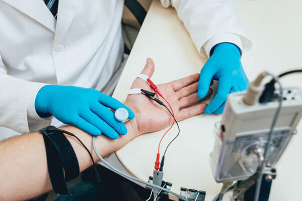
You may need to do special tests to get more information about the nerve. One common test is the nerve conduction velocity (NCV) test. The NCV test measures the speed of the impulses traveling along the nerve. Impulses are slowed when the nerve is compressed or constricted.
The NCV test is sometimes combined with an electromyogram (EMG). The EMG tests the muscles of the forearm that are controlled by the ulnar nerve to see whether the muscles are working properly. If they aren’t, it may be because the nerve is not working well.
Radial tunnel syndrome happens when the radial nerve is squeezed where it passes through a tunnel near the elbow. The symptoms of radial tunnel syndrome are very similar to the symptoms of tennis elbow (lateral epicondylitis). There are very few helpful tests for radial tunnel syndrome, which can make it hard to diagnose.
What is the radial tunnel?
The radial nerve starts at the side of the neck, where the individual nerve roots leave the spine. The nerve roots exit through small openings between the vertebrae. These openings are called neural foramina.
The nerve roots join together to form three main nerves that travel down the arm to the hand. One of these nerves is the radial nerve. The nerve passes down the back of the upper arm. It then spirals outward and crosses the outside (the lateral part) of the elbow before it winds its way down the forearm and hand.
On the lateral part of the elbow, the radial nerve enters a tunnel formed by muscles and bone. This is called the radial tunnel.
Passing through the radial tunnel, the radial nerve runs below the supinator muscle. The supinator muscle lets you twist your right hand clockwise. This is the motion of using a screwdriver to tighten a screw.
After the radial nerve passes under the supinator muscle, it branches out and attaches to the muscles on the back of the forearm.
Causes
What causes the pain of radial tunnel syndrome?
Pain is caused by pressure on the radial nerve. There are several spots along the radial tunnel that can pinch the nerve. If the tunnel is too small, it can squeeze the nerve and cause pain. Repetitive, forceful pushing and pulling, bending of the wrist, gripping, and pinching can also stretch and irritate the nerve.
Sometimes a direct blow to the outside of the elbow can injure the radial nerve. Constant twisting movements of the arm, common in assembly work, can also pinch the radial nerve and lead to radial tunnel syndrome.
Symptoms
What does radial tunnel syndrome feel like?
The symptoms of radial tunnel syndrome are tenderness and pain on the outside of the elbow. The symptoms of radial tunnel syndrome are very similar to the symptoms of tennis elbow. As in tennis elbow, pain from radial tunnel syndrome often starts near the lateral epicondyle. (The lateral epicondyle is a bony point on the outside of your elbow joint.) The pain gets worse when you bend your wrist backward, turn your palm upward, or hold something with a stiff wrist or straightened elbow.
One difference between radial tunnel syndrome and tennis elbow is the exact location of the pain. In tennis elbow, the pain starts where the tendon attaches to the lateral epicondyle. In radial tunnel syndrome, the pain is centered about two inches further down the arm, over the spot where the radial nerve goes under the supinator muscle. Radial tunnel syndrome may also cause a more achy type of pain or fatigue in the muscles of the forearm. Nerve pressure inside the radial tunnel leads to weakness in the muscles on the back of the forearm and wrist, making it difficult to steady the wrist when grasping and lifting. It can even lead to wrist drop, meaning the back of the hand can’t be cocked up. Skin sensation is not changed because the sensory portion of the radial nerve branches off above the elbow and does not enter the radial tunnel
Diagnosis
How will my doctor know I have radial tunnel syndrome?
The diagnosis of radial tunnel syndrome can be difficult. Many cases are initially diagnosed as tennis elbow. Tests don’t always help tell the two conditions apart.
Your doctor will take a detailed medical history. You will be asked questions about your pain, your activities, and any past injuries to your elbow.
Your doctor will then do a physical examination to look for the most painful spot. The prodding and movement may hurt. But it is important that your doctor know exactly where and when you have pain. Pinpointing the source of the pain will be most helpful in determining whether you have radial tunnel syndrome or tennis elbow.
You may do some tests of the radial nerve. An electromyogram (EMG) tests to see if the muscles of the forearm are working properly. If the test shows a problem with the muscles, it may be caused by a problem with the radial nerve. The nerve conduction velocity (NCV) test measures the speed of an electrical impulse as it travels along the radial nerve. If the speed is too slow, then the nerve is probably pinched.
These tests are not very accurate in diagnosing radial tunnel syndrome. Many people who have radial tunnel syndrome will have normal EMG and NCV test results. Your doctor will consider all parts of the examination in diagnosing whether or not you have a problem with radial tunnel syndrome.
Olecranon bursitis is inflammation of a small sac of fluid located on the tip of the elbow. This inflammation can cause many problems in the elbow.
Anatomy
Where is the olecranon bursa, and what does it do?
A bursa is a sac made of thin, slippery tissue. Bursae occur in the body wherever skin, muscles, or tendons need to slide over bone. Bursae are lubricated with a small amount of fluid inside that helps reduce friction from the sliding parts.
The olecranon bursa is located between the tip, or point, of the elbow (called the olecranon) and the overlying skin. This bursa allows the elbow to bend and straighten freely underneath the skin.
Causes
How does olecranon bursitis develop?
Bursitis is the inflammation of a bursa. The olecranon bursa can become irritated and inflamed in a number of ways.
In some cases, a direct blow or a fall onto the elbow can damage the bursa. This usually causes bleeding into the bursa sac, because the blood vessels in the tissues that make up the bursa are damaged and torn. In the skin this would simply form a bruise, but in a bursa blood may actually fill the bursa sac. This causes the bursa to swell up like a rubber balloon filled with water.
The blood in the bursa is thought to cause an inflammatory reaction. The walls of the bursa may thicken and remain thickened and tender even after the blood has been absorbed by the body. This thickening and swelling of the bursa is referred to as olecranon bursitis.
Olecranon bursitis can also occur over a longer period of time. People who constantly put their elbows on a hard surface as part of their activities or job can repeatedly injure the bursa. This repeated injury can lead to irritation and thickening of the bursa over time. The chronic irritation leads to the same condition in the end: olecranon bursitis.
The olecranon bursa can also become infected. This may occur without any warning, or it may be caused by a small injury and infection of the skin over the bursa that spreads down into the bursa. In this case, instead of blood or inflammatory fluid in the bursa, it becomes filled with pus. The area around the bursa becomes hot, red, and very tender.
Symptoms
What does olecranon bursitis feel like?
Olecranon bursitis causes pain and swelling in the area at the tip of the elbow. It may be very difficult to put the elbow down on a surface due to the tenderness. If the condition has been present for some time, small lumps may be felt underneath the skin over the olecranon. Sometimes these lumps feel as though something is floating around in the olecranon bursa, and they can be very tender. These lumps are usually the thickened folds of bursa tissue that have formed in response to chronic inflammation.
The bursa sac may swell and fill with fluid at times. This is usually related to your activity level, and more activity usually causes more swelling. Over time the bursa can grow very thick, almost like an elbow pad on the olecranon.
Finally, if the bursa becomes infected, the elbow becomes swollen and very tender and warm to the touch around the bursa. You may run a fever and feel chills. An abscess, or area of pus, may form on the elbow. If the infection is not treated quickly, the abscess may even begin to drain, meaning the pus begins to seep out.
Diagnosis
How do doctors identify the condition?
The diagnosis of olecranon bursitis is usually obvious from the physical examination. In cases where the elbow swells immediately after a fall or other injury to the elbow, X-rays may be necessary to make sure that the elbow isn’t fractured. Usually chronic olecranon bursitis is also easy to diagnose without any special tests.
If your doctor is uncertain whether or not the bursa is infected, a needle may be inserted into the bursa and the fluid removed. This fluid will be sent to a lab for tests. The results are used to determine whether infection is present. If so, the type of bacteria that is causing the infection is identified. Your doctor will use this information to know what antibiotic will work best to cure the infection.
Golfers elbow is a relatively common overuse injury which typically causes pain at the inner aspect of the elbow. It is a similar condition to tennis elbow however affects the inner aspect of the elbow rather than the outer aspect.
The group of muscles at the front of the forearm are commonly called the forearm flexors (figure 1). These muscles act to flex the wrist and fingers (i.e. bend them forwards) and have a common bony attachment at the inner aspect of the elbow called the medial epicondyle (figure 1). The forearm flexors attach to the medial epicondyle via the flexor tendon.
During contraction of the forearm flexors, tension is placed through the flexor tendon at its attachment to the medial epicondyle. When this tension is excessive due to too much repetition or high force, damage to the tendon occurs. Golfers elbow is a condition whereby there is damage, with subsequent inflammation and degeneration to the flexor tendon at its bony attachment to the inner elbow. This is usually due to gradual wear and tear associated with overuse, however, it may also occur traumatically due to a specific incident.
Although this condition can occur at any age, it is commonly seen in patients between the ages of 40 and 60.
Causes of golfers elbow
Contrary to what the name suggests, you do not have to play golf to develop this condition. In fact, golfers elbow is more commonly seen in non-golf players than in golf players. Patients typically develop this condition due to activities involving repetitive wrist flexion against resistance or forceful or repetitive gripping of the hand. These activities may include sports or manual work such as:
- golf (especially those who continually take divots out of the ground)
- tennis (especially those players who put a lot of top spin on the ball)
- squash
- badminton
- water skiing
- gymnastics
- body building or weight lifting
- carpentry
- hammering
- painting
- chopping wood
- bricklaying
- repetitive use of a screwdriver
- sewing
- knitting
- working at a computer
It is also common for patients to develop this condition following a sudden increase in activities that place stress on the forearm flexors (such as involvement in a golf tournament over consecutive days) or due to a change in these activities (such as using a new technique or clubs, or hitting the ball too hard). In golf players, golfers elbow is often associated with poor swing technique.
Occasionally, this condition may develop suddenly. This is usually due to a forceful movement involving a heavy lifting or gripping force through the arm. In golf, this may occur when mis-timing a shot and taking a divot out of hard ground.
A history of wrist, elbow, shoulder or neck injury may increase the likelihood of a patient developing this condition.
Signs and symptoms of golfers elbow
The symptoms associated with golfers elbow usually develop gradually over a period of time. Initially, symptoms may present as an ache following an aggravating or unaccustomed activity. This may often be felt first thing in the morning. Patients with this condition usually experience localized elbow pain 1-2cm down from the bony lump on the inner aspect of the elbow (medial epicondyle – figure 1) that increases on firmly touching this region. Occasionally, the pain may radiate into the forearm.
In less severe cases of this condition, patients may only experience a minor ache. In more severe cases, the pain may be quite incapacitating and can keep the patient awake at night. Usually pain is experienced as an ache that increases to a sharper pain with activity. Occasionally, golfers elbow can be associated with neck, shoulder or upper back pain on the same side. In longstanding cases muscle weakness and reduced grip strength may also be present.
Patients with this condition often experience an increase in pain during everyday activities such as picking up a cup, turning a door knob, opening a jar, shaking hands, carrying groceries or turning the steering wheel of a car. Elbow stiffness may also be experienced and is typically worse first thing in the morning.
Diagnosis of golfers elbow
A thorough subjective and objective examination from a physiotherapist is usually sufficient to diagnose golfers elbow. Further investigations such as an MRI scan or Ultrasound may be required, in rare cases, to confirm diagnosis
Tennis elbow is a common overuse injury which typically causes pain at the outer aspect of the elbow. Although it can occur at any age, it is commonly seen in patients between the ages of 40 and 50.
The group of muscles at the back of the forearm are commonly called the forearm extensors (figure 1). These muscles act to extend the wrist and fingers (i.e. bend the wrist backwards and straighten the fingers) and have a common bony attachment at the outer aspect of the elbow called the lateral epicondyle (figure 1). The forearm extensors attach to the lateral epicondyle via the extensor tendon.
During contraction of the forearm extensors, tension is placed through the extensor tendon at its attachment to the lateral epicondyle. When this tension is excessive due to too much repetition or high force, damage to the tendon occurs. Tennis elbow is a condition whereby there is damage, with subsequent inflammation and degeneration of the extensor tendon at its bony attachment to the outer elbow. It usually occurs due to gradual wear and tear associated with overuse, however, the condition may also occur traumatically due to a specific incident. The extensor muscle that is most commonly affected in this condition is known as the extensor carpi radialis brevis.
Causes of tennis elbow
Contrary to what the name suggests, you do not have to play tennis to develop this condition. In fact, tennis elbow is more commonly seen in non-tennis players than in tennis players. Patients typically develop this condition in association with activities involving repeated wrist extension against resistance. This includes sporting activities such as tennis, squash, badminton, as well as manual work such as carpentry, painting, chopping wood, bricklaying, repetitive use of a screwdriver, sewing and knitting or working at a computer. Patients may also develop this condition from other activities involving repetitive or forceful gripping of the hand.
It is common for patients to develop this condition following a sudden increase in activities that place stress on the forearm extensors (such as involvement in a tennis tournament over consecutive days) or due to a change in these activities (such as hitting balls in the wet, hitting into a strong breeze, tightening the tennis racquet’s strings, using a new tennis racquet or technique, or simply hitting the ball too hard). Occasionally, the condition may develop suddenly. This is usually due to a forceful movement involving a heavy lifting or gripping force through the arm. In tennis players, tennis elbow is often associated with poor backhand technique. A history of wrist, elbow, shoulder or neck injury may also increase the likelihood of developing this condition.
Signs and symptoms of tennis elbow
The symptoms associated with this condition usually develop gradually over a period of time. Initially, symptoms may present as an ache following an aggravating or unaccustomed activity. This may often be felt first thing in the morning. Patients usually experience localized elbow pain 1-2cm down from the bony lump on the outer aspect of the elbow (lateral epicondyle – figure 1) that increases on firmly touching this region. Occasionally, pain may radiate into the forearm.
In less severe cases of this condition, patients may only experience a minor ache. In more severe cases, pain may be quite incapacitating and can keep the patient awake at night. Usually pain is experienced as an ache that increases to a sharper pain with activity. Occasionally, this condition can be associated with neck or upper back pain on the same side. In longstanding cases muscle weakness and reduced grip strength may also be present.
Patients with tennis elbow often experience an increase in pain during everyday activities such as gripping activities, picking up a cup, turning a door knob, opening a jar, shaking hands, carrying groceries or turning the steering wheel of a car. Elbow stiffness may also be experienced and is typically worse first thing in the morning.
Diagnosis of tennis elbow
A thorough subjective and objective examination from a physiotherapist is usually sufficient to diagnose tennis elbow. Further investigations such as an MRI scan or Ultrasound may be required, in rare cases, to confirm diagnosis.
When the joint surfaces of an elbow are forced apart, the elbow is dislocated. The elbow is the second most commonly dislocated joint in adults (after shoulder dislocation). Elbow dislocations can be complete or partial. A partial dislocation is referred to as a subluxation. The amount of force needed to cause an elbow dislocation is enough to cause a bone fracture at the same time. These two injuries (dislocation-fracture) often occur together.
What part of the elbow is affected?
The bones of the elbow are the humerus (the upper arm bone), the ulna (the larger bone of the forearm, on the opposite side of the thumb), and the radius (the smaller bone of the forearm on the same side as the thumb).
The elbow itself is essentially a hinge joint, meaning it bends and straightens like a hinge. But there is a second joint where the end of the radius (the radial head) meets the humerus. This joint is complicated because the radius has to rotate so that you can turn your hand palm up and palm down. At the same time, it has to slide against the end of the humerus as the elbow bends and straightens. The joint is even more complex because the radius has to slide against the ulna as it rotates the wrist as well. As a result, the end of the radius at the elbow is shaped like a smooth knob with a cup at the end to fit on the end of the humerus. The edges are also smooth where it glides against the ulna.
Articular cartilage is the material that covers the ends of the bones of any joint. Articular cartilage can be up to one-quarter of an inch thick in the large, weight-bearing joints. It is a bit thinner in joints such as the elbow, which don’t support weight. Articular cartilage is white, shiny, and has a rubbery consistency. It is slippery, which allows the joint surfaces to slide against one another without causing any damage. In the elbow, articular cartilage covers the end of the humerus, the end of the radius, and the end of the ulna.
There are several important ligaments in the elbow. Ligaments are soft tissue structures that connect bones to bones. The ligaments around a joint usually combine together to form a joint capsule. A joint capsule is a watertight sac that surrounds a joint and contains lubricating fluid called synovial fluid.
In the elbow, two of the most important ligaments are themedial collateral ligament and the lateral collateral ligament. The medial collateral is on the inside edge of the elbow, and the lateral collateral is on the outside edge. Together these two ligaments connect the humerus to the ulna and keep it tightly in place as it slides through the groove at the end of the humerus. These ligaments are the main source of stability for the elbow. They can be torn when there is an injury to or dislocation of the elbow. If they do not heal correctly the elbow can be too loose, or unstable.
There is also an important ligament called the annular ligament that wraps around the radial head and holds it tight against the ulna. The word annular means ring-shaped. The annular ligament forms a ring around the radial head as it holds it in place. This ligament can be torn when the entire elbow or just the radial head is dislocated.
Causes
What can cause this condition?
Elbow dislocation is often the result of trauma. The most common trauma resulting in an elbow dislocation is a fall onto an outstretched hand and arm. When the hand hits the ground, the force is transmitted through the forearm to the elbow. This force pushes the elbow out of its socket.
This can also result in a fracture/dislocation. About half of all elbow dislocations in teens and young adults occur as a result of a sports activity. The most common elbow dislocations are associated with sports such as gymnastics, cycling, rollerblading, or skateboarding.
Dislocation can also occur from a sideswipe injury. This type of injury occurs when the driver of an automobile has the elbow out the open window during a car accident. The force of the impact causes a severe fracture-dislocation of the elbow.
Symptoms
What does this condition feel like?
If the elbow is fully dislocated, it will look out of joint. There may be dimples or indentations of the skin over the dislocation where the bones have shifted position. Pain can be intense until the arm is relocated. The pain is often relieved immediately after the joint is put back in place. There may be some residual tenderness around the joint.
If ligaments or other soft tissues are torn, there can be swelling and bruising around the elbow. Bruising is not immediately obvious but appears several days after the injury. Injury to any of the three nerves that cross the elbow (median, ulnar, radial nerves) can cause neurologic symptoms such as numbness, tingling, and/or weakness of the forearm, wrist, and hand. If a bone fracture is also involved the fracture can cut or damage a nerve causing temporary or permanent paralysis.
Pain and an inability to straighten the elbow or pain when turning the hand the palm up (supination) is typical. There is often tenderness along the lateral aspect of the elbow (side of the elbow away from the body).
Diagnosis
How do doctors diagnose elbow dislocation?
The history and physical examination are probably the most important tools the physician uses to guide his or her diagnosis. Moving the elbow passively is painful, especially extension and supination. The doctor will check for any signs of injury to the nerves or blood vessels.
X-ray is the best way to look for dislocation or fracture-dislocation.
After the joint is relocated, other imaging studies may be ordered to look for damage to the joint cartilage, bone, ligaments, and other soft tissues. If bone detail is difficult to identify on an X-ray, a computed tomography (CT) scan may be done. If it is important to evaluate the ligaments, a magnetic resonance image (MRI) can be helpful.
A frozen shoulder is a condition characterized by inflammation, scarring and tightening of the connective tissue surrounding the shoulder joint, usually resulting in shoulder pain and a marked loss of shoulder movement.
The shoulder joint is a ball and socket joint. The shoulder blade gives rise to the socket of the shoulder, whilst the ball arises from the top of the humerus (upper arm bone). Surrounding the ball and socket joint is strong connective tissue holding the bones together known as the shoulder joint capsule (figure 1).
Occasionally, the shoulder joint capsule may become inflamed with subsequent tightening and scarring of the shoulder joint capsule. When this occurs the condition is known as a frozen shoulder and usually results in a marked loss of shoulder range of movement.
Frozen shoulders most commonly occur in people over 40 years of age and typically affect women more commonly than men. They can generally be divided into 3 phases, each of which can last a number of months:
- Pain – In this first phase of a frozen shoulder, the shoulder typically becomes painful with most movements. The shoulder may also start to stiffen during this phase.
- Freezing – This second phase of this condition is characterized by a marked loss of movement of the shoulder, coinciding with scarring of the shoulder joint capsule. Patients typically experience difficulty when elevating the arm or taking their hand behind their back. Pain may decrease noticeably during this phase.
- Thawing – In this final phase, the shoulder spontaneously begins to ‘loosen’ up and movement to the shoulder is gradually restored.
Causes of a frozen shoulder
Whilst the exact cause of a frozen shoulder is not exactly known, it is thought to occur following injury or damage to the shoulder joint or adjacent soft tissue. In these cases, a frozen shoulder is more likely to develop if the initial injury is not treated appropriately. This often occurs due to inadequate rest from aggravating activities or adopting a ‘no pain no gain’ attitude. A patient may also have an increased likelihood of developing the condition following excessive immobility (i.e. not moving the shoulder, particularly after injury or surgery) or if they suffer from other auto-immune diseases or diabetes.
Signs and symptoms of a frozen shoulder
The symptoms associated with this condition usually develop gradually over time (often following a recent history of shoulder injury, surgery and/or immobilisation). Patients typically experience a dull ache that may increase to a sharper pain with certain movements or activities. Pain tends to be focused deep in the shoulder, however may occasionally be experienced in the upper arm, upper back and neck. Patients may also experience stiffness in each of these regions.
The pain associated with a frozen shoulder may increase with any movement of the shoulder and with activities placing stress on the shoulder joint. These activities may include: arm elevation, lifting, carrying, pushing or pulling, lying on the affected side and taking your hand behind your back (e.g. putting on a bra). Patients with a frozen shoulder often experience pain at night or upon waking in the morning. As the condition progresses from the painful phase to the frozen phase, pain may reduce significantly.
Aside from pain, patients also typically experience marked stiffness and significantly reduced range of movement of the shoulder. This typically affects all shoulder movements, but is most noticeable with rotation and elevation. This may present as difficulty elevating the affected arm or an inability to take the hand behind the back. As this condition progresses from the frozen phase to the thawing phase, range of movement gradually increases with a subsequent reduction in joint stiffness. Patients may also develop muscle wasting in the affected arm, as the condition progresses, through lack of use.
Although a frozen shoulder generally affects only one side, some patients may develop the condition in both shoulders.
Diagnosis of a frozen shoulder
A thorough subjective and objective examination from a physiotherapist is usually sufficient to diagnose a frozen shoulder. Further investigations such as an Ultrasound or MRI scan may be required to assist diagnosis.
Subacromial bursitis is a condition characterized by tissue damage and inflammation of the subacromial bursa (a small fluid filled sac located beneath the bony prominence at the top / outer aspect of the shoulder) causing pain in the shoulder.
The shoulder joint is a ball and socket joint. The socket arises from the outer aspect of the shoulder blade, whilst the ball arises from the upper aspect of the humerus (upper arm bone). Just above the ball and socket joint of the shoulder is a bony prominence known as the acromion .
Beneath the acromion lies a bursa known as the subacromial bursa (figure 1). A bursa is a small sac filled with lubricating fluid and is designed to reduce friction between adjacent soft tissue or bony layers. The subacromial bursa reduces friction between the bony prominence of the acromion (above the bursa) and the tendon of the supraspinatus muscle (which attaches to the upper aspect of the humeral head) below the bursa.
During certain activities, such as arm elevation, rotating the shoulder, lifting, pushing or pulling or lying on the shoulder, friction and compressive forces are placed on the subacromial bursa. Pressure may also be placed on the subacromial bursa following a direct impact or fall onto the point of the shoulder, elbow or outstretched hand. When these forces are excessive due to too much repetition or high force, irritation and inflammation of the bursa may occur. When this occurs, the condition is known as subacromial bursitis.
Subacromial bursitis often occurs in association with other conditions of the shoulder such as a rotator cuff tear or tendinopathy, shoulder impingement or shoulder instability.
Causes of subacromial bursitis
Subacromial bursitis most commonly occurs due to repetitive or prolonged activities placing strain on the subacromial bursa. This typically occurs due to:
- repetitive or prolonged overhead activities
- repetitive or prolonged arm elevation activities
- repetitive or prolonged use of the arm in front of the body
- activities involving rotation of the shoulder
- lifting (especially overhead)
- excessive pushing or pulling activities (placing strain on the bursa via the supraspinatus tendon)
- putting weight through the affected arm
- lying on the affected side
Occasionally, the condition may occur suddenly due a direct blow to the point of the shoulder or due to a fall onto the shoulder, elbow or outstretched hand.
In athletes, subacromial bursitis may be seen in throwing sports (such as cricket or baseball), swimming (particularly freestyle and butterfly), racquet sports (such as tennis), weight lifting or paddling sports (such as kayaking).
Anomalies of the acromion bone, degenerative changes to the AC joint, or bony spurs known as osteophytes, may predispose a patient to developing subacromial bursitis.
Signs and symptoms of subacromial bursitis
Patients with this condition typically experience pain at the top, front, back or outer aspect of the shoulder. Pain may also radiate into the upper arm as far as the elbow. In less severe cases, patients may only experience an ache or stiffness in the shoulder that increases with rest following activities placing strain on the bursa. These activities typically include arm elevation activities, use of the arm in front of the body or overhead, shoulder rotating activities, lifting, pushing or pulling, placing weight through the arm or lying on the affected side. The pain associated with this condition may also warm up with activity in the initial stages of injury.
As the condition progresses, patients may experience symptoms that increase during activity or sport, affecting performance. Patients with subacromial bursitis may also experience pain on firmly touching the top / outer aspect of the shoulder. A painful arc of arm elevation and / or a feeling of shoulder weakness may also be present particularly when attempting to lift or elevate the arm overhead.
Diagnosis of subacromial bursitis
A thorough subjective and objective examination from a physiotherapist may be sufficient to diagnose subacromial bursitis. An ultrasound is usually the common investigation used to identify subacromial bursitis. Further investigations such as an X-ray, CT or MRI scan are often required to assist diagnosis and assess the severity of the condition or the presence of other conditions.
Introduction
A distal biceps rupture occurs when the tendon attaching the biceps muscle to the elbow is torn from the bone. This injury occurs mainly in middle-aged men during heavy work or lifting. A distal biceps rupture is rare compared to ruptures where the top of the biceps connects at the shoulder. Distal biceps ruptures make up only three percent of all biceps tendon ruptures.
Anatomy
What parts of the elbow are affected?
The biceps muscle goes from the shoulder to the elbow on the front of the upper arm. Tendons attach muscles to bone. Two separate tendons connect the upper part of the biceps muscle to the shoulder. One tendon connects the lower end of the biceps to the elbow.
The lower biceps tendon is called the distal biceps tendon. The word distal means that the tendon is further down the arm. The upper two tendons of the biceps are called the proximal biceps tendons, because they are closer to the top of the arm.
The distal biceps tendon attaches to a small bump on the radius bone of the forearm. This small bony bump is called the radial tuberosity. The radius is the smaller of the two bones between the elbow and the wrist that make up the forearm. The radius goes from the outside edge of the elbow to the thumb side of the wrist. It parallels the larger bone of the forearm, the ulna. The ulna goes from the inside edge of the elbow to the wrist.
The upper (proximal) end of the biceps has two attachments to the shoulder. The main attachment is the long head of the biceps. It connects the biceps muscle to the top of the shoulder socket, called the glenoid. The short head of the biceps angles up and in to its attachment on the corocoid process of the shoulder blade. The corocoid process is a small bony knob in the front of the shoulder.
Tendons are made up of strands of a material called collagen. The collagen strands are lined up in bundles next to each other.
Because the collagen strands in tendons are lined up, tendons have high tensile strength. This means they can withstand high forces that pull on both ends of the tendon. When muscles contract, they pull on one end of the tendon. The other end of the tendon pulls on the bone, causing the bone to move.
Contracting the biceps muscle can bend the elbow upward. The biceps can also help flex the shoulder, raising the arm up. And the biceps can rotate, or twist, the forearm in a way that points the palm of the hand up. This movement is called supination. Supination positions the hand as if you were carrying a tray.
Causes
Why did I develop a rupture of the distal biceps?
The most common cause of a distal biceps rupture happens when a middle-aged man lifts a box or other heavy item with his elbows bent. Often the load is heavier than expected, or the load may shift unexpectedly during the lift. This forces the elbow to straighten, even though the biceps muscle is working hard to keep the elbow bent. The biceps muscle contracts extra hard to help handle the load. As tension on the muscle and tendon increases, the distal biceps tendon snaps or tears where it connects to the radius.
Symptoms
What does a ruptured distal biceps feel like?
When the distal biceps tendon ruptures, it usually sounds and feels like a pop directly in front of the elbow. At first the pain is intense. The pain often subsides quickly after a complete rupture because tension is immediately taken off the pain sensors in the tendon. Swelling and bruising in front of the elbow usually develop shortly after the pop. The biceps may appear to have balled up near the elbow. The arm often feels weak with attempts to bend the elbow, lift the shoulder, or twist the forearm into supination (palm up).
The distal biceps tendon sometimes tears only part of the way. When this happens, a pop may not be felt or heard. Instead, the area in front of the elbow may simply be painful, and the arm may feel weak with the same arm movements that are affected in a complete rupture.
Diagnosis
How can my doctor be sure I have ruptured the distal biceps?
An early diagnosis is best. If surgery is needed, people who’ve ruptured their distal biceps tendon usually have better results when surgery is done soon after the injury.
Your doctor will first take a detailed medical history. You will need to answer questions about your pain, how your pain affects you, your regular activities, and past injuries to your elbow.
The physical exam is often most helpful in diagnosing a rupture of the distal biceps tendon. Your doctor may position your elbow and forearm to see which movements are painful and weak. By feeling the muscle and tendon, your doctor can often tell if the tendon has ruptured off the bone.
X-rays may be ordered. X-rays are mainly used to find out if there are other injuries in the elbow. Plain X-rays do not show soft tissues like tendons. They will not show a distal biceps rupture unless a small piece of bone got pulled off the radius as the tendon ruptured. This type of injury is called an avulsion fracture.
Your doctor may also order a magnetic resonance imaging (MRI) scan to see if the biceps tendon is only partially torn or if the tendon fully ruptured. An MRI is a special imaging test that uses magnetic waves to create pictures of the elbow in slices. The MRI can also show if there are other problems in the elbow.
Diseases
-
Neck Pain
-
Shoulder & Elbow
-
Hand & Wrist
-
Arthritis
-
Lower Back Pain
-
Hip & Femur
-
Knee & Lower Leg
-
Foot & Ankle
-
Osteoporosis