Arthritis
Arthritis literally means “inflammation of a joint.” In some forms of arthritis, such as osteoarthritis, the inflammation arises because the smooth covering (articular cartilage) on the ends of bones become damaged or worn. Osteoarthritis is usually found in one, usually weightbearing, joint.
In other forms of arthritis, such as rheumatoid arthritis, the joint lining becomes inflamed as part of a disease process that affects the entire body. Some other types of arthritis are: seronegative spondyloarthropathies, crytalline deposition diseases, and septic arthritis.
Arthritis is a major cause of lost work time and serious disability for many people. Although arthritis is mainly a disease of adults, children may also have it.
Anatomy
Arthritis is a disease of the joint. A joint is where the ends of two or more bones meet. The knee joint, for example, is formed between the bones of the lower leg (the tibia and the fibula) and the thighbone (the femur). The hip joint is where the top of the thighbone (femoral head) meets a concave portion of the pelvis (the acetabulum).
A smooth tissue of cartilage covers the ends of bones in a joint. Cartilage cushions the bone and allows the joint to move easily without the friction that would come with bone-on-bone contact. A joint is enclosed by a fibrous envelope, called the synovium, which produces a fluid that also helps to reduce friction and wear in a joint. Ligaments connect the bones and keep the joint stable. Muscles and tendons power the joint and enable it to move.
There are two major categories of arthritis.
The first type is caused by wear and tear on the articular cartilage (osteoarthritis) through the natural aging process, through constant use, or through trauma (post-traumatic arthritis).
The second type is caused by one of a number of inflammatory processes.
Regardless of whether the cause is from injury, normal wear and tear, or disease, the joint becomes inflamed, causing swelling, pain and stiffness. This is usually temporary. Inflammation is one of the body’s normal reactions to injury or disease. In arthritic joints, however, inflammation may cause long-lasting or permanent disability.
Arthritis of the Foot and Ankle
Arthritis is the leading cause of disability in the United States. It can occur at any age, and literally means “pain within a joint.” As a result, arthritis is a term used broadly to refer to a number of different conditions.
Although there is no cure for arthritis, there are many treatment options available. It is important to seek help early so that treatment can begin as soon as possible. With treatment, people with arthritis are able to manage pain, stay active, and live fulfilling lives, often without surgery.
Description
There are three types of arthritis that may affect your foot and ankle.
Osteoarthritis
Osteoarthritis, also known as degenerative or “wear and tear” arthritis, is a common problem for many people after they reach middle age. Over the years, the smooth, gliding surface covering the ends of bones (cartilage) becomes worn and frayed. This results in inflammation, swelling, and pain in the joint.
Osteoarthritis progresses slowly and the pain and stiffness it causes worsens over time.
Rheumatoid Arthritis
Unlike osteoarthritis which follows a predictable pattern in certain joints, rheumatoid arthritis is a system-wide disease. It is an inflammatory disease where the patient’s own immune system attacks and destroys cartilage.
Post-Traumatic Arthritis
Post-traumatic arthritis can develop after an injury to the foot or ankle. This type of arthritis is similar to osteoarthritis and may develop years after a fracture, severe sprain, or ligament injury.
Cause
Osteoarthritis
Many factors increase your risk for developing osteoarthritis. Because the ability of cartilage to heal itself decreases as we age, older people are more likely to develop the disease. Other risk factors include obesity and family history of the disease.
Rheumatoid Arthritis
The exact cause of rheumatoid arthritis is not known. Although it is not an inherited disease, researchers believe that some people have genes that make them more susceptible. There is usually a “trigger,” such as an infection or environmental factor, which activates the genes. When the body is exposed to this trigger, the immune system begins to produce substances that attack the joint. This is what may lead to the development of rheumatoid arthritis.
Post-Traumatic Arthritis
Fractures – particularly those that damage the joint surface – and dislocations are the most common injuries that lead to this type of arthritis. An injured joint is about seven times more likely to become arthritic, even if the injury is properly treated. In fact, following injury, your body can secrete hormones that stimulate the death of your cartilage cells.
Anatomy
There are 28 bones and more than 30 joints in the foot. Tough bands of tissue, called ligaments, keep the bones and joints in place. If arthritis develops in one or more of these joints, balance and walking may be affected.
Joints and bones of the foot and ankle.
The joints most commonly affected by arthritis in the lower extremity include:
- The ankle (tibiotalar joint). The ankle is where the shinbone (tibia) rests on the uppermost bone of the foot (the talus).
- The three joints of the hindfoot. These three joints include:
- The subtalar or talocalcaneal joint, where the bottom of the talusconnects to the heel bone (calcaneus);
- The talonavicular joint, where the talus connects to the inner midfoot bone (navicular); and
- The calcaneocuboid joint, where the heel bone connects to the outer midfoot bone (cuboid).
- The midfoot (metatarsocuneiform joint). This is where one of the forefoot bones (metatarsals) connects to the smaller midfoot bones (cuneiforms).
- The great toe (first metatarsophalangeal joint). This is where the first metatarsal connects to the great toe bone (phalange).This is also the area where bunions usually develop.
Symptoms
Signs and symptoms of arthritis of the foot vary, depending on which joint is affected. Common symptoms include:
- Pain or tenderness
- Stiffness or reduced motion
- Swelling
- Difficulty walking due to any of the above
What is knee arthritis?
Knee arthritis is a condition characterized by a degenerative process whereby there is gradual wear and tear to the cartilage and bone surfaces of the knee joint with subsequent inflammation. Knee arthritis may occur following a specific injury or due to repetitive forces going through the knee joint beyond what it can withstand over a period of time.
In a normal knee, joint surfaces are smooth and there is cartilage between the bone ends which allows for efficient shock absorption and smooth movement (figure 1).
When the knee is damaged or overloaded, particularly with excessive weight-bearing or twisting forces, degeneration of the cartilage can occur, reducing the knee’s shock absorbing capacity. As the condition progresses, and the cartilage wears away, the joint space can narrow and there is eventual wearing down of the bone ends so that the surfaces are no longer smooth (figure 2). The bone ends may also develop small bony processes (spurs) called ‘osteophytes’. When some or all of these changes occur, the condition is known as knee arthritis.
Arthritis of the knee usually occurs after the age of 50 years and tends to affect females more frequently than males. It is more common in those patients who are overweight or have a past history of injury, surgery or trauma to the knee.
Signs and symptoms of knee arthritis
Patients with this condition usually experience symptoms that develop gradually over time. In patients with minor cases of knee arthritis, little or no symptoms may be present. As the condition progresses, there may be increasing knee pain with weight bearing activity and joint stiffness (particularly after rest or first thing in the morning). Swelling, decreased flexibility (i.e. an inability to fully straighten or bend the knee), severe joint pain, pain at night and a grinding, clicking or locking sensation during certain movements may also be experienced. Symptoms can sometimes fluctuate from month to month with patients reporting an increase in symptoms with colder weather. In more severe cases, muscle wasting (especially of the quadriceps), a visible deformity of the knee joint, and a limp may also be present.
Diagnosis of knee arthritis
A thorough subjective and objective examination from a physiotherapist may be sufficient to diagnose knee arthritis. An X-ray is usually required to confirm diagnosis and may demonstrate signs of decreased joint space, irregularities of the bony ends and/or the presence of bony spurs (osteophytes). Sometimes an MRI may also be indicated to assist with diagnosis and rule out other pathologies.
Arthritis of the Shoulder
In 2011, more than 50 million people in the United States reported that they had been diagnosed with some form of arthritis, according to the National Health Interview Survey. Simply defined, arthritis is inflammation of one or more of your joints. In a diseased shoulder, inflammation causes pain and stiffness.
Although there is no cure for arthritis of the shoulder, there are many treatment options available. Using these, most people with arthritis are able to manage pain and stay active.
Your shoulder is made up of three bones: your upper arm bone (humerus), your shoulder blade (scapula), and your collarbone (clavicle).
The head of your upper arm bone fits into a rounded socket in your shoulder blade. This socket is called the glenoid. A combination of muscles and tendons keeps your arm bone centered in your shoulder socket. These tissues are called the rotator cuff.
There are two joints in the shoulder, and both may be affected by arthritis. One joint is located where the clavicle meets the tip of the shoulder blade (acromion). This is called the acromioclavicular (AC) joint.
Where the head of the humerus fits into the scapula is called the glenohumeral joint.
To provide you with effective treatment, your physician will need to determine which joint is affected and what type of arthritis you have.
Description
Five major types of arthritis typically affect the shoulder.
Osteoarthritis
Also known as “wear-and-tear” arthritis, osteoarthritis is a condition that destroys the smooth outer covering (articular cartilage) of bone. As the cartilage wears away, it becomes frayed and rough, and the protective space between the bones decreases. During movement, the bones of the joint rub against each other, causing pain.
Osteoarthritis usually affects people over 50 years of age and is more common in the acromioclavicular joint than in the glenohumeral shoulder joint.
Rheumatoid Arthritis
Rheumatoid arthritis (RA) is a chronic disease that attacks multiple joints throughout the body. It is symmetrical, meaning that it usually affects the same joint on both sides of the body.
The joints of your body are covered with a lining — called synovium — that lubricates the joint and makes it easier to move. Rheumatoid arthritis causes the lining to swell, which causes pain and stiffness in the joint.
Rheumatoid arthritis is an autoimmune disease. This means that the immune system attacks its own tissues. In RA, the defenses that protect the body from infection instead damage normal tissue (such as cartilage and ligaments) and soften bone.
Rheumatoid arthritis is equally common in both joints of the shoulder.
Posttraumatic Arthritis
Posttraumatic arthritis is a form of osteoarthritis that develops after an injury, such as a fracture or dislocation of the shoulder.
Rotator Cuff Tear Arthropathy
Arthritis can also develop after a large, long-standing rotator cuff tendon tear. The torn rotator cuff can no longer hold the head of the humerus in the glenoid socket, and the humerus can move upward and rub against the acromion. This can damage the surfaces of the bones, causing arthritis to develop.
The combination of a large rotator cuff tear and advanced arthritis can lead to severe pain and weakness, and the patient may not be able to lift the arm away from the side.
Avascular Necrosis
Avascular necrosis (AVN) of the shoulder is a painful condition that occurs when the blood supply to the head of the humerus is disrupted. Because bone cells die without a blood supply, AVN can ultimately lead to destruction of the shoulder joint and arthritis.
Avascular necrosis develops in stages. As it progresses, the dead bone gradually collapses, which damages the articular cartilage covering the bone and leads to arthritis. At first, AVN affects only the head of the humerus, but as AVN progresses, the collapsed head of the humerus can damage the glenoid socket.
Causes of AVN include high dose steroid use, heavy alcohol consumption, sickle cell disease, and traumatic injury, such as fractures of the shoulder. In some cases, no cause can be identified; this is referred to as idiopathic AVN
Arthritis of the Hand
The hand and wrist have multiple small joints that work together to produce motion, including the fine motion needed to thread a needle or tie a shoelace. When the joints are affected by arthritis, activities of daily living can be difficult. Arthritis can occur in many areas of the hand and wrist and can have more than one cause.
Over time, if the arthritis is not treated, the bones that make up the joint can lose their normal shape. This causes more pain and further limits motion.
Description
Simply defined, arthritis is inflammation of one or more of your joints. The most common types of arthritis are osteoarthritis and rheumatoid arthritis, but there are more than 100 different forms.
Healthy joints move easily because of a smooth, slippery tissue called articular cartilage. Cartilage covers the ends of bones and provides a smooth gliding surface for the joint. This smooth surface is lubricated by a fluid that looks and feels like oil. It is produced by the joint lining called synovium.
Disease
When arthritis occurs due to disease, the onset of symptoms is gradual and the cartilage decreases slowly. The two most common forms of arthritis from disease are osteoarthritis and rheumatoid arthritis.
Osteoarthritis is much more common and generally affects older people. Also known as “wear and tear” arthritis, osteoarthritis causes cartilage to wear away. It appears in a predictable pattern in certain joints.
Rheumatoid arthritis is a chronic disease that can affect many parts of your body. It causes the joint lining (synovium) to swell, which causes pain and stiffness in the joint. Rheumatoid arthritis most often starts in the small joints of the hands and feet. It usually affects the same joints on both sides of the body.
Trauma
Fractures, particularly those that damage the joint surface, and dislocations are among the most common injuries that lead to arthritis. Even when properly treated, an injured joint is more likely to become arthritic over time.
Fractures within the finger joints.
Symptoms
Pain
Early symptoms of arthritis of the hand include joint pain that may feel “dull,” or a “burning” sensation. The pain often occurs after periods of increased joint use, such as heavy gripping or grasping. The pain may not be present immediately, but may show up hours later or even the following day. Morning pain and stiffness are typical.
As the cartilage wears away and there is less material to provide shock absorption, the symptoms occur more frequently. In advanced disease, the joint pain may wake you up at night.
Pain might be made worse with use and relieved by rest. Many people with arthritis complain of increased joint pain with rainy weather. Activities that once were easy, such as opening a jar or starting the car, become difficult due to pain. To prevent pain at the arthritic joint, you might change the way you use your hand.
Swelling
When the affected joint is subject to greater stress than it can bear, it may swell in an attempt to prevent further joint use.
Changes in Surrounding Joints
In patients with advanced thumb base arthritis, the neighboring joints may become more mobile than normal.
Warmth
The arthritic joint may feel warm to touch. This is due to the body’s inflammatory response.
Crepitation and Looseness
There may be a sensation of grating or grinding in the affected joint (crepitation). This is caused by damaged cartilage surfaces rubbing against one another. If arthritis is due to damaged ligaments, the support structures of the joint may be unstable or “loose.” In advanced cases, the joint may appear larger than normal (hypertrophic). This is usually due to a combination of bone changes, loss of cartilage, and joint swelling.
Cysts
When arthritis affects the end joints of the fingers (DIP joints), small cysts (mucous cysts) may develop. The cysts may then cause ridging or dents in the nail plate of the affected finger.
Arthritis of the Thumb
Arthritis is a condition that irritates or destroys a joint. Although there are several types of arthritis, the one that most often affects the joint at the base of the thumb (the basal joint) is osteoarthritis (degenerative or “wear-and-tear” arthritis).
Description
Smooth cartilage covers the ends of the bones. It enables the bones to glide easily in the joint. Without it, bones rub against each other, causing friction and damage to the bones and the joint. Osteoarthritis occurs when the cartilage begins to wear away.
The joint at the base of the thumb, near the wrist and at the fleshy part of the thumb, enables the thumb to swivel, pivot, and pinch so that you can grip things in your hand.
Arthritis of the base of the thumb is more common in women than in men, and usually occurs after 40 years of age. Prior fractures or other injuries to the joint may increase the likelihood of developing this condition.
Symptoms
- Pain with activities that involve gripping or pinching, such as turning a key, opening a door, or snapping your fingers
- Swelling and tenderness at the base of the thumb
- An aching discomfort after prolonged use
- Loss of strength in gripping or pinching activities
- An enlarged, “out-of-joint” appearance
- Development of a bony prominence or bump over the joint
- Limited motion
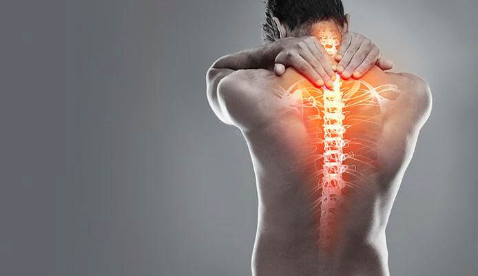
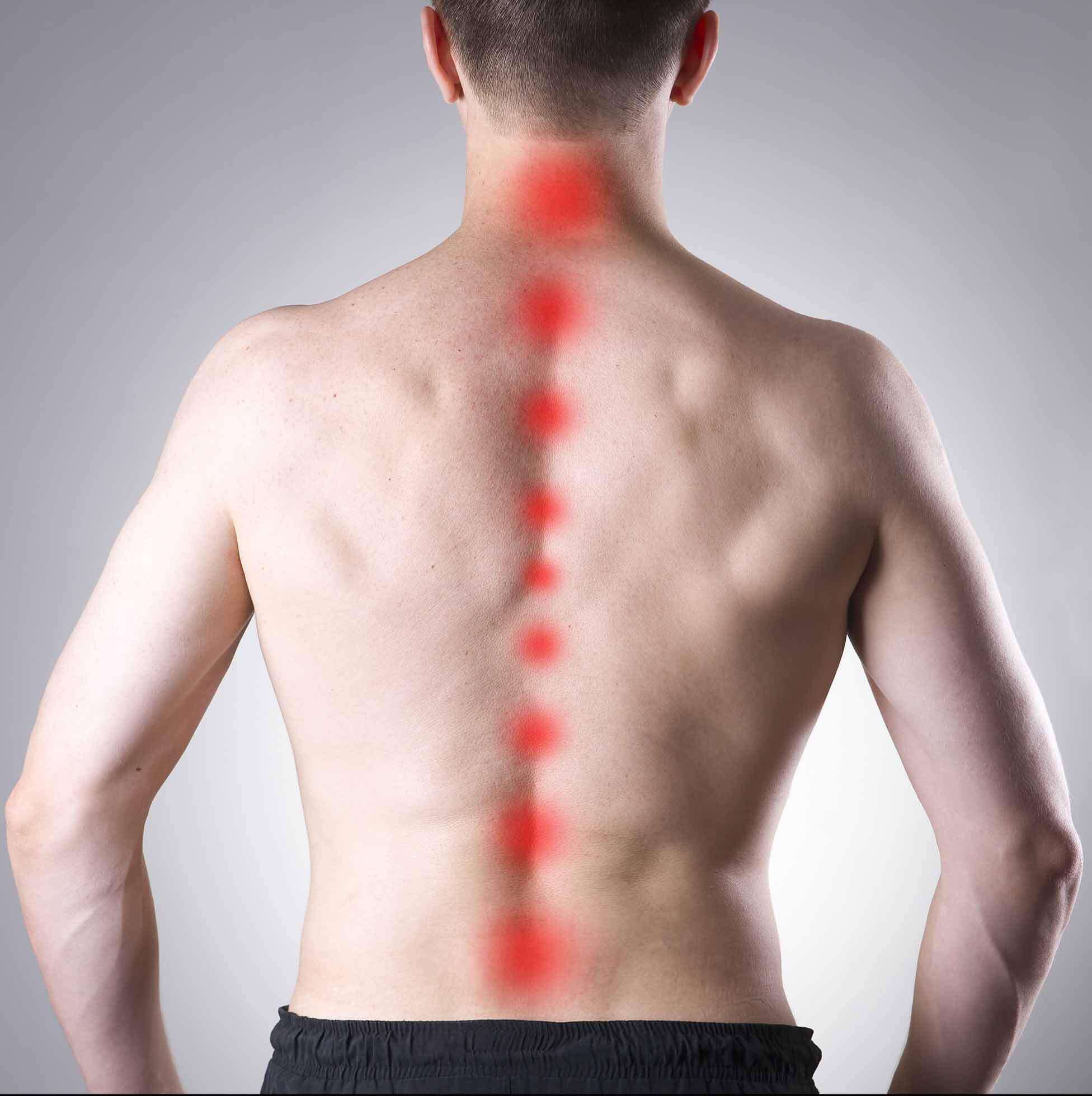
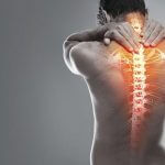
Arthritis of the lumbar facet joints can be a source of significant low back pain. Aligned on the back of the spinal column, the facet joints link each vertebra together. Articular cartilage covers the surfaces where these joints meet. Like other joints in the body that are covered with articular cartilage, the lumbar facet joints can be affected by arthritis.
What part of the back is involved?
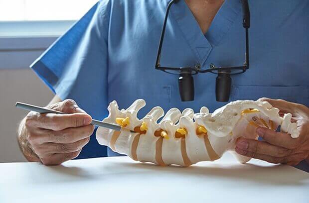
The human spine is made up of 24 spinal bones, called vertebrae. Vertebrae are stacked on top of one another to create the spinal column. The spinal column gives the body its form. It is the body’s main upright support.
The back portion of the spinal column forms a bony ring. When the vertebrae are stacked on top of each other, these bony rings create a hollow tube. This tube, called the spinal canal, surrounds the spinal cord as it passes through the spine. Just as the skull protects the brain, the bones of the spinal column protect the spinal cord.
Between the vertebrae of each spinal segment are two facet joints. The facet joints are located on the back of the spinal column. There are two facet joints between each pair of vertebrae, one on each side of the spine. A facet joint is made of small, bony knobs that line up along the back of the spine. Where these knobs meet, they form a joint that connects the two vertebrae. The alignment of the facet joints of the lumbar spine allows freedom of movement as you bend forward and back.
The surfaces of the facet joints are covered by articular cartilage. Articular cartilage is a smooth, rubbery material that covers the ends of most joints. It allows the bone ends to move against each other smoothly, without friction.
Why do I have this problem?
Normally, the facet joints fit together snugly and glide smoothly, without pressure. If pressure builds where the joint meets, the cartilage on the joint surfaces wears off, or erodes.
Each segment in the spine has three main points of movement, the intervertebral disc and the two facet joints. Injury or problems in any one of these structures affects the other two. As a disc thins with aging and from daily wear and tear, the space between two spinal vertebrae shrinks. This causes the facet joints to press together.
Facet joints can also become arthritic due to a back injury earlier in life. Fractures, torn ligaments, and disc problems can all cause abnormal movement and alignment, putting extra stress on the surfaces of the facet joints.
The body responds to this extra pressure by developing bone spurs. As the spurs form around the edges of the facet joints, the joints become enlarged. This is called hypertrophy. Eventually, the joint surfaces become arthritic. When the articular cartilage degenerates, or wears away, the bone underneath is uncovered and rubs against bone. The joint becomes inflamed, swollen, and painful.
Facet joint arthritis develops slowly over a long period of time. This is partly because spinal degeneration in later life is the main cause of facet joint arthritis. Symptoms rarely develop immediately when degeneration is causing the problems. However, rapid movements, heavy twisting, or backward motions in the low back can injure a facet joint, leading to immediate symptoms.
Symptoms
Pain from facet joint arthritis is usually worse after resting or sleeping. Also, bending the trunk sideways or backward usually produces pain on the same side as the arthritic facet joint. For example, if you lie on your stomach on a flat surface and raise your upper body, you hyperextend the spine. This increases pressure on the facet joints and can cause pain if there is facet joint arthritis.
Pain may be felt in the center of the low back and can spread into one or both buttocks. Sometimes the pain spreads into the thighs, but it rarely goes below the knee. Numbness and tingling, the symptoms of nerve compression, are usually not felt because facet arthritis generally causes only mechanical pain. Mechanical pain comes from abnormal movement in the spine.
However, symptoms of nerve compression can sometimes occur at the same time as the facet joint pain. The arthritis can cause bone spurs at the edges of the facet joint. These bone spurs may form in the opening where the nerve root leaves the spinal canal. This opening is called the neural foramen. If the bone spurs rub against the nerve root, the nerve can become inflamed and irritated. This nerve irritation can cause symptoms where the nerve travels. These symptoms may include numbness, tingling, slowed reflexes, and muscle weakness.
Diseases
-
Neck Pain
-
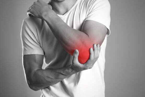
Shoulder & Elbow
-
Hand & Wrist
-
Arthritis
-
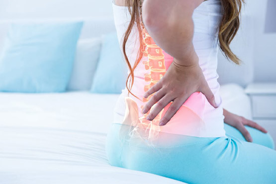
Lower Back Pain
-
Hip & Femur
-
Knee & Lower Leg
-
Foot & Ankle
-
Osteoporosis